Arsenic »
PDB 1b92-1fyx »
1c82 »
Arsenic in PDB 1c82: Mechanism of Hyaluronan Binding and Degradation: Structure of Streptococcus Pneumoniae Hyaluronate Lyase in Complex with Hyaluronic Acid Disaccharide at 1.7 A Resolution
Enzymatic activity of Mechanism of Hyaluronan Binding and Degradation: Structure of Streptococcus Pneumoniae Hyaluronate Lyase in Complex with Hyaluronic Acid Disaccharide at 1.7 A Resolution
All present enzymatic activity of Mechanism of Hyaluronan Binding and Degradation: Structure of Streptococcus Pneumoniae Hyaluronate Lyase in Complex with Hyaluronic Acid Disaccharide at 1.7 A Resolution:
4.2.2.1;
4.2.2.1;
Protein crystallography data
The structure of Mechanism of Hyaluronan Binding and Degradation: Structure of Streptococcus Pneumoniae Hyaluronate Lyase in Complex with Hyaluronic Acid Disaccharide at 1.7 A Resolution, PDB code: 1c82
was solved by
K.Ponnuraj,
M.J.Jedrzejas,
with X-Ray Crystallography technique. A brief refinement statistics is given in the table below:
| Resolution Low / High (Å) | 40.00 / 1.70 |
| Space group | P 21 21 21 |
| Cell size a, b, c (Å), α, β, γ (°) | 83.550, 104.190, 99.880, 90.00, 90.00, 90.00 |
| R / Rfree (%) | 21.5 / 25.7 |
Other elements in 1c82:
The structure of Mechanism of Hyaluronan Binding and Degradation: Structure of Streptococcus Pneumoniae Hyaluronate Lyase in Complex with Hyaluronic Acid Disaccharide at 1.7 A Resolution also contains other interesting chemical elements:
| Sodium | (Na) | 2 atoms |
Arsenic Binding Sites:
The binding sites of Arsenic atom in the Mechanism of Hyaluronan Binding and Degradation: Structure of Streptococcus Pneumoniae Hyaluronate Lyase in Complex with Hyaluronic Acid Disaccharide at 1.7 A Resolution
(pdb code 1c82). This binding sites where shown within
5.0 Angstroms radius around Arsenic atom.
In total 2 binding sites of Arsenic where determined in the Mechanism of Hyaluronan Binding and Degradation: Structure of Streptococcus Pneumoniae Hyaluronate Lyase in Complex with Hyaluronic Acid Disaccharide at 1.7 A Resolution, PDB code: 1c82:
Jump to Arsenic binding site number: 1; 2;
In total 2 binding sites of Arsenic where determined in the Mechanism of Hyaluronan Binding and Degradation: Structure of Streptococcus Pneumoniae Hyaluronate Lyase in Complex with Hyaluronic Acid Disaccharide at 1.7 A Resolution, PDB code: 1c82:
Jump to Arsenic binding site number: 1; 2;
Arsenic binding site 1 out of 2 in 1c82
Go back to
Arsenic binding site 1 out
of 2 in the Mechanism of Hyaluronan Binding and Degradation: Structure of Streptococcus Pneumoniae Hyaluronate Lyase in Complex with Hyaluronic Acid Disaccharide at 1.7 A Resolution
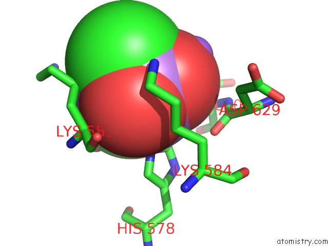
Mono view
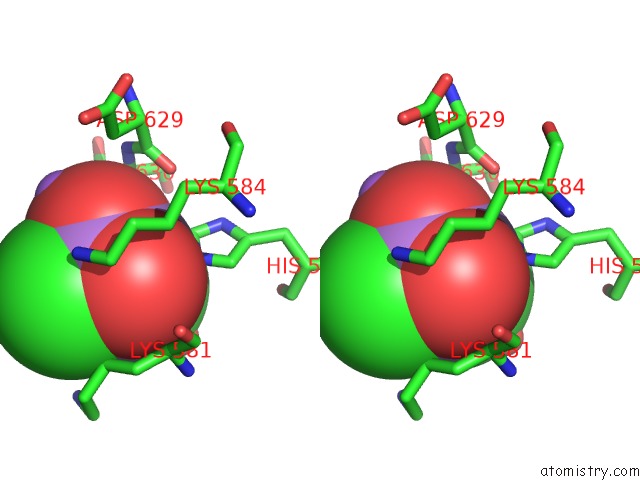
Stereo pair view
Mono view
Stereo pair view
A full contact list of Arsenic with other atoms in the As binding
site number 1 of Mechanism of Hyaluronan Binding and Degradation: Structure of Streptococcus Pneumoniae Hyaluronate Lyase in Complex with Hyaluronic Acid Disaccharide at 1.7 A Resolution within 5.0Å range:
|
Arsenic binding site 2 out of 2 in 1c82
Go back to
Arsenic binding site 2 out
of 2 in the Mechanism of Hyaluronan Binding and Degradation: Structure of Streptococcus Pneumoniae Hyaluronate Lyase in Complex with Hyaluronic Acid Disaccharide at 1.7 A Resolution
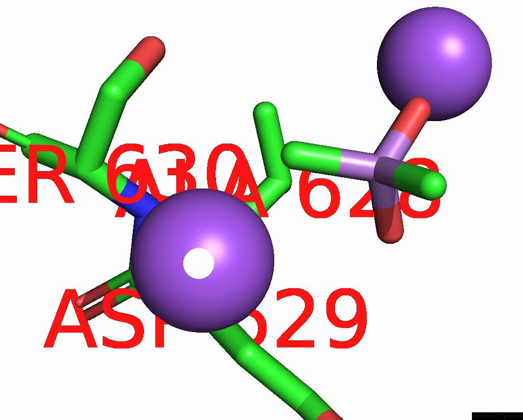
Mono view
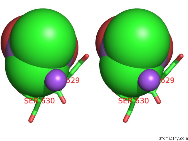
Stereo pair view
Mono view
Stereo pair view
A full contact list of Arsenic with other atoms in the As binding
site number 2 of Mechanism of Hyaluronan Binding and Degradation: Structure of Streptococcus Pneumoniae Hyaluronate Lyase in Complex with Hyaluronic Acid Disaccharide at 1.7 A Resolution within 5.0Å range:
|
Reference:
K.Ponnuraj,
M.J.Jedrzejas.
Mechanism of Hyaluronan Binding and Degradation: Structure of Streptococcus Pneumoniae Hyaluronate Lyase in Complex with Hyaluronic Acid Disaccharide at 1.7 A Resolution. J.Mol.Biol. V. 299 885 2000.
ISSN: ISSN 0022-2836
PubMed: 10843845
DOI: 10.1006/JMBI.2000.3817
Page generated: Wed Jul 10 10:58:17 2024
ISSN: ISSN 0022-2836
PubMed: 10843845
DOI: 10.1006/JMBI.2000.3817
Last articles
Zn in 9MJ5Zn in 9HNW
Zn in 9G0L
Zn in 9FNE
Zn in 9DZN
Zn in 9E0I
Zn in 9D32
Zn in 9DAK
Zn in 8ZXC
Zn in 8ZUF