Arsenic »
PDB 1glj-1pqu »
1glj »
Arsenic in PDB 1glj: Escherichia Coli Glycerol Kinase Mutant with Bound Atp Analog Showing Substantial Domain Motion
Enzymatic activity of Escherichia Coli Glycerol Kinase Mutant with Bound Atp Analog Showing Substantial Domain Motion
All present enzymatic activity of Escherichia Coli Glycerol Kinase Mutant with Bound Atp Analog Showing Substantial Domain Motion:
2.7.1.30;
2.7.1.30;
Protein crystallography data
The structure of Escherichia Coli Glycerol Kinase Mutant with Bound Atp Analog Showing Substantial Domain Motion, PDB code: 1glj
was solved by
C.E.Bystrom,
D.W.Pettigrew,
B.P.Branchaud,
S.J.Remington,
with X-Ray Crystallography technique. A brief refinement statistics is given in the table below:
| Resolution Low / High (Å) | 20.00 / 3.00 |
| Space group | C 2 2 21 |
| Cell size a, b, c (Å), α, β, γ (°) | 99.668, 200.699, 114.733, 90.00, 90.00, 90.00 |
| R / Rfree (%) | n/a / n/a |
Other elements in 1glj:
The structure of Escherichia Coli Glycerol Kinase Mutant with Bound Atp Analog Showing Substantial Domain Motion also contains other interesting chemical elements:
| Magnesium | (Mg) | 1 atom |
Arsenic Binding Sites:
The binding sites of Arsenic atom in the Escherichia Coli Glycerol Kinase Mutant with Bound Atp Analog Showing Substantial Domain Motion
(pdb code 1glj). This binding sites where shown within
5.0 Angstroms radius around Arsenic atom.
In total 2 binding sites of Arsenic where determined in the Escherichia Coli Glycerol Kinase Mutant with Bound Atp Analog Showing Substantial Domain Motion, PDB code: 1glj:
Jump to Arsenic binding site number: 1; 2;
In total 2 binding sites of Arsenic where determined in the Escherichia Coli Glycerol Kinase Mutant with Bound Atp Analog Showing Substantial Domain Motion, PDB code: 1glj:
Jump to Arsenic binding site number: 1; 2;
Arsenic binding site 1 out of 2 in 1glj
Go back to
Arsenic binding site 1 out
of 2 in the Escherichia Coli Glycerol Kinase Mutant with Bound Atp Analog Showing Substantial Domain Motion
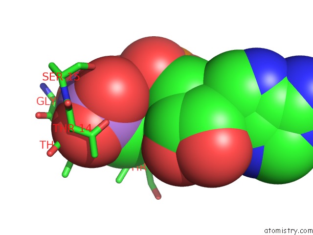
Mono view
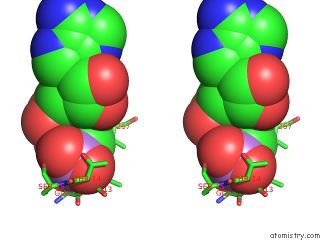
Stereo pair view
Mono view
Stereo pair view
A full contact list of Arsenic with other atoms in the As binding
site number 1 of Escherichia Coli Glycerol Kinase Mutant with Bound Atp Analog Showing Substantial Domain Motion within 5.0Å range:
|
Arsenic binding site 2 out of 2 in 1glj
Go back to
Arsenic binding site 2 out
of 2 in the Escherichia Coli Glycerol Kinase Mutant with Bound Atp Analog Showing Substantial Domain Motion
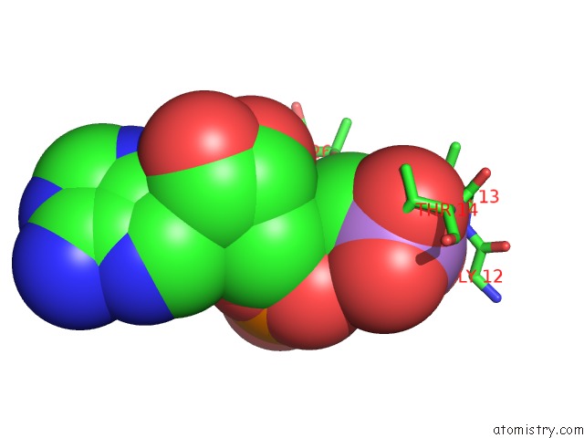
Mono view
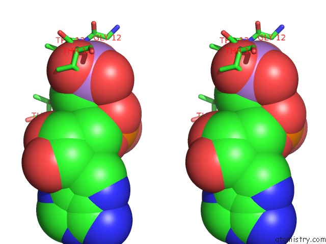
Stereo pair view
Mono view
Stereo pair view
A full contact list of Arsenic with other atoms in the As binding
site number 2 of Escherichia Coli Glycerol Kinase Mutant with Bound Atp Analog Showing Substantial Domain Motion within 5.0Å range:
|
Reference:
C.E.Bystrom,
D.W.Pettigrew,
B.P.Branchaud,
P.O'brien,
S.J.Remington.
Crystal Structures of Escherichia Coli Glycerol Kinase Variant S58-->W in Complex with Nonhydrolyzable Atp Analogues Reveal A Putative Active Conformation of the Enzyme As A Result of Domain Motion. Biochemistry V. 38 3508 1999.
ISSN: ISSN 0006-2960
PubMed: 10090737
DOI: 10.1021/BI982460Z
Page generated: Sun Jul 6 22:55:09 2025
ISSN: ISSN 0006-2960
PubMed: 10090737
DOI: 10.1021/BI982460Z
Last articles
Fe in 2YXOFe in 2YRS
Fe in 2YXC
Fe in 2YNM
Fe in 2YVJ
Fe in 2YP1
Fe in 2YU2
Fe in 2YU1
Fe in 2YQB
Fe in 2YOO