Arsenic »
PDB 1glj-1pqu »
1ihu »
Arsenic in PDB 1ihu: Crystal Structure of the Escherichia Coli Arsenite-Translocating Atpase in Complex with Mg-Adp-ALF3
Enzymatic activity of Crystal Structure of the Escherichia Coli Arsenite-Translocating Atpase in Complex with Mg-Adp-ALF3
All present enzymatic activity of Crystal Structure of the Escherichia Coli Arsenite-Translocating Atpase in Complex with Mg-Adp-ALF3:
3.6.3.16;
3.6.3.16;
Protein crystallography data
The structure of Crystal Structure of the Escherichia Coli Arsenite-Translocating Atpase in Complex with Mg-Adp-ALF3, PDB code: 1ihu
was solved by
T.Zhou,
S.Radaev,
B.P.Rosen,
D.L.Gatti,
with X-Ray Crystallography technique. A brief refinement statistics is given in the table below:
| Resolution Low / High (Å) | 25.15 / 2.15 |
| Space group | I 2 2 2 |
| Cell size a, b, c (Å), α, β, γ (°) | 73.897, 75.945, 222.607, 90.00, 90.00, 90.00 |
| R / Rfree (%) | 21.5 / 26.2 |
Other elements in 1ihu:
The structure of Crystal Structure of the Escherichia Coli Arsenite-Translocating Atpase in Complex with Mg-Adp-ALF3 also contains other interesting chemical elements:
| Fluorine | (F) | 3 atoms |
| Magnesium | (Mg) | 2 atoms |
| Aluminium | (Al) | 1 atom |
| Cadmium | (Cd) | 8 atoms |
| Chlorine | (Cl) | 3 atoms |
Arsenic Binding Sites:
The binding sites of Arsenic atom in the Crystal Structure of the Escherichia Coli Arsenite-Translocating Atpase in Complex with Mg-Adp-ALF3
(pdb code 1ihu). This binding sites where shown within
5.0 Angstroms radius around Arsenic atom.
In total only one binding site of Arsenic was determined in the Crystal Structure of the Escherichia Coli Arsenite-Translocating Atpase in Complex with Mg-Adp-ALF3, PDB code: 1ihu:
In total only one binding site of Arsenic was determined in the Crystal Structure of the Escherichia Coli Arsenite-Translocating Atpase in Complex with Mg-Adp-ALF3, PDB code: 1ihu:
Arsenic binding site 1 out of 1 in 1ihu
Go back to
Arsenic binding site 1 out
of 1 in the Crystal Structure of the Escherichia Coli Arsenite-Translocating Atpase in Complex with Mg-Adp-ALF3
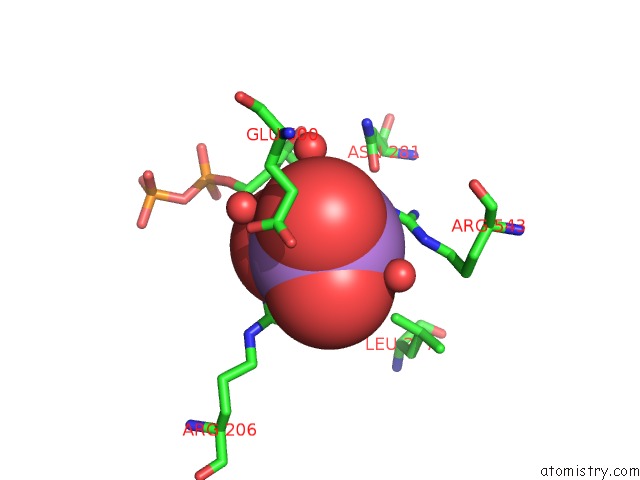
Mono view
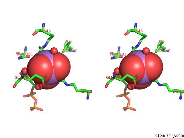
Stereo pair view
Mono view
Stereo pair view
A full contact list of Arsenic with other atoms in the As binding
site number 1 of Crystal Structure of the Escherichia Coli Arsenite-Translocating Atpase in Complex with Mg-Adp-ALF3 within 5.0Å range:
|
Reference:
T.Zhou,
S.Radaev,
B.P.Rosen,
D.L.Gatti.
Conformational Changes in Four Regions of the Escherichia Coli Arsa Atpase Link Atp Hydrolysis to Ion Translocation. J.Biol.Chem. V. 276 30414 2001.
ISSN: ISSN 0021-9258
PubMed: 11395509
DOI: 10.1074/JBC.M103671200
Page generated: Sun Jul 6 22:55:55 2025
ISSN: ISSN 0021-9258
PubMed: 11395509
DOI: 10.1074/JBC.M103671200
Last articles
Ca in 2YEQCa in 2YG0
Ca in 2YFZ
Ca in 2YFS
Ca in 2YFU
Ca in 2YFT
Ca in 2YDP
Ca in 2YFR
Ca in 2YC4
Ca in 2YDX