Arsenic »
PDB 1glj-1pqu »
1pqu »
Arsenic in PDB 1pqu: Crystal Structure of the H277N Mutant of Aspartate Semialdehyde Dehydrogenase From Haemophilus Influenzae Bound with Nadp, S-Methyl Cysteine Sulfoxide and Cacodylate
Enzymatic activity of Crystal Structure of the H277N Mutant of Aspartate Semialdehyde Dehydrogenase From Haemophilus Influenzae Bound with Nadp, S-Methyl Cysteine Sulfoxide and Cacodylate
All present enzymatic activity of Crystal Structure of the H277N Mutant of Aspartate Semialdehyde Dehydrogenase From Haemophilus Influenzae Bound with Nadp, S-Methyl Cysteine Sulfoxide and Cacodylate:
1.2.1.11;
1.2.1.11;
Protein crystallography data
The structure of Crystal Structure of the H277N Mutant of Aspartate Semialdehyde Dehydrogenase From Haemophilus Influenzae Bound with Nadp, S-Methyl Cysteine Sulfoxide and Cacodylate, PDB code: 1pqu
was solved by
J.Blanco,
R.A.Moore,
R.E.Viola,
with X-Ray Crystallography technique. A brief refinement statistics is given in the table below:
| Resolution Low / High (Å) | 44.65 / 1.92 |
| Space group | P 1 21 1 |
| Cell size a, b, c (Å), α, β, γ (°) | 72.929, 76.579, 134.097, 90.00, 92.59, 90.00 |
| R / Rfree (%) | 18.2 / 23.4 |
Arsenic Binding Sites:
The binding sites of Arsenic atom in the Crystal Structure of the H277N Mutant of Aspartate Semialdehyde Dehydrogenase From Haemophilus Influenzae Bound with Nadp, S-Methyl Cysteine Sulfoxide and Cacodylate
(pdb code 1pqu). This binding sites where shown within
5.0 Angstroms radius around Arsenic atom.
In total 4 binding sites of Arsenic where determined in the Crystal Structure of the H277N Mutant of Aspartate Semialdehyde Dehydrogenase From Haemophilus Influenzae Bound with Nadp, S-Methyl Cysteine Sulfoxide and Cacodylate, PDB code: 1pqu:
Jump to Arsenic binding site number: 1; 2; 3; 4;
In total 4 binding sites of Arsenic where determined in the Crystal Structure of the H277N Mutant of Aspartate Semialdehyde Dehydrogenase From Haemophilus Influenzae Bound with Nadp, S-Methyl Cysteine Sulfoxide and Cacodylate, PDB code: 1pqu:
Jump to Arsenic binding site number: 1; 2; 3; 4;
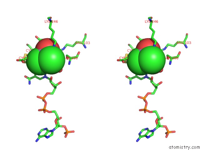
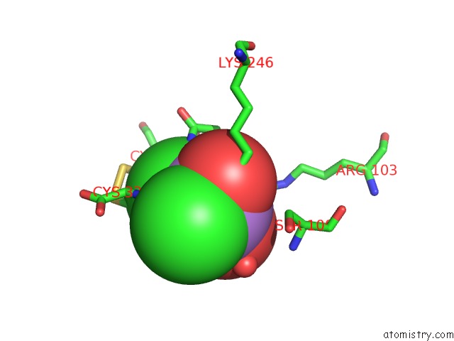
Arsenic binding site 1 out of 4 in 1pqu
Go back to
Arsenic binding site 1 out
of 4 in the Crystal Structure of the H277N Mutant of Aspartate Semialdehyde Dehydrogenase From Haemophilus Influenzae Bound with Nadp, S-Methyl Cysteine Sulfoxide and Cacodylate

Mono view

Stereo pair view
Mono view
Stereo pair view
A full contact list of Arsenic with other atoms in the As binding
site number 1 of Crystal Structure of the H277N Mutant of Aspartate Semialdehyde Dehydrogenase From Haemophilus Influenzae Bound with Nadp, S-Methyl Cysteine Sulfoxide and Cacodylate within 5.0Å range:
|
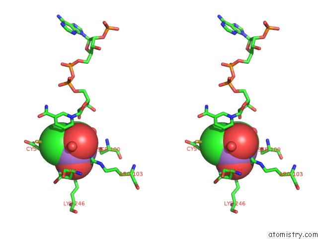
Arsenic binding site 2 out of 4 in 1pqu
Go back to
Arsenic binding site 2 out
of 4 in the Crystal Structure of the H277N Mutant of Aspartate Semialdehyde Dehydrogenase From Haemophilus Influenzae Bound with Nadp, S-Methyl Cysteine Sulfoxide and Cacodylate

Mono view
Stereo pair view
Mono view
Stereo pair view
A full contact list of Arsenic with other atoms in the As binding
site number 2 of Crystal Structure of the H277N Mutant of Aspartate Semialdehyde Dehydrogenase From Haemophilus Influenzae Bound with Nadp, S-Methyl Cysteine Sulfoxide and Cacodylate within 5.0Å range:
|
Arsenic binding site 3 out of 4 in 1pqu
Go back to
Arsenic binding site 3 out
of 4 in the Crystal Structure of the H277N Mutant of Aspartate Semialdehyde Dehydrogenase From Haemophilus Influenzae Bound with Nadp, S-Methyl Cysteine Sulfoxide and Cacodylate
Mono view

Stereo pair view
Mono view
Stereo pair view
A full contact list of Arsenic with other atoms in the As binding
site number 3 of Crystal Structure of the H277N Mutant of Aspartate Semialdehyde Dehydrogenase From Haemophilus Influenzae Bound with Nadp, S-Methyl Cysteine Sulfoxide and Cacodylate within 5.0Å range:
|
Arsenic binding site 4 out of 4 in 1pqu
Go back to
Arsenic binding site 4 out
of 4 in the Crystal Structure of the H277N Mutant of Aspartate Semialdehyde Dehydrogenase From Haemophilus Influenzae Bound with Nadp, S-Methyl Cysteine Sulfoxide and Cacodylate

Mono view
Stereo pair view
Mono view
Stereo pair view
A full contact list of Arsenic with other atoms in the As binding
site number 4 of Crystal Structure of the H277N Mutant of Aspartate Semialdehyde Dehydrogenase From Haemophilus Influenzae Bound with Nadp, S-Methyl Cysteine Sulfoxide and Cacodylate within 5.0Å range:
|
Reference:
J.Blanco,
R.A.Moore,
C.R.Faehnle,
D.M.Coe,
R.E.Viola.
The Role of Substrate-Binding Groups in the Mechanism of Aspartate-Beta-Semialdehyde Dehydrogenase. Acta Crystallogr.,Sect.D V. 60 1388 2004.
ISSN: ISSN 0907-4449
PubMed: 15272161
DOI: 10.1107/S0907444904012971
Page generated: Wed Jul 10 11:10:41 2024
ISSN: ISSN 0907-4449
PubMed: 15272161
DOI: 10.1107/S0907444904012971
Last articles
Zn in 9JYWZn in 9IR4
Zn in 9IR3
Zn in 9GMX
Zn in 9GMW
Zn in 9JEJ
Zn in 9ERF
Zn in 9ERE
Zn in 9EGV
Zn in 9EGW