Arsenic »
PDB 1q2o-1yhc »
1y0r »
Arsenic in PDB 1y0r: Crystal Structure of the Tetrahedral Aminopeptidase From P. Horikoshii
Protein crystallography data
The structure of Crystal Structure of the Tetrahedral Aminopeptidase From P. Horikoshii, PDB code: 1y0r
was solved by
M.Groll,
L.Borissenko,
with X-Ray Crystallography technique. A brief refinement statistics is given in the table below:
| Resolution Low / High (Å) | 14.91 / 1.75 |
| Space group | P 2 3 |
| Cell size a, b, c (Å), α, β, γ (°) | 111.572, 111.572, 111.572, 90.00, 90.00, 90.00 |
| R / Rfree (%) | 21 / 22.7 |
Other elements in 1y0r:
The structure of Crystal Structure of the Tetrahedral Aminopeptidase From P. Horikoshii also contains other interesting chemical elements:
| Zinc | (Zn) | 2 atoms |
Arsenic Binding Sites:
The binding sites of Arsenic atom in the Crystal Structure of the Tetrahedral Aminopeptidase From P. Horikoshii
(pdb code 1y0r). This binding sites where shown within
5.0 Angstroms radius around Arsenic atom.
In total only one binding site of Arsenic was determined in the Crystal Structure of the Tetrahedral Aminopeptidase From P. Horikoshii, PDB code: 1y0r:
In total only one binding site of Arsenic was determined in the Crystal Structure of the Tetrahedral Aminopeptidase From P. Horikoshii, PDB code: 1y0r:
Arsenic binding site 1 out of 1 in 1y0r
Go back to
Arsenic binding site 1 out
of 1 in the Crystal Structure of the Tetrahedral Aminopeptidase From P. Horikoshii
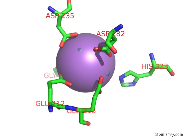
Mono view
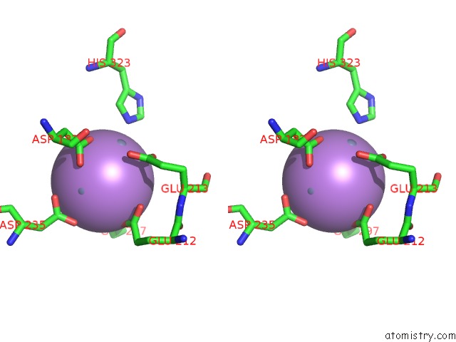
Stereo pair view
Mono view
Stereo pair view
A full contact list of Arsenic with other atoms in the As binding
site number 1 of Crystal Structure of the Tetrahedral Aminopeptidase From P. Horikoshii within 5.0Å range:
|
Reference:
L.Borissenko,
M.Groll.
Crystal Structure of Tet Protease Reveals Complementary Protein Degradation Pathways in Prokaryotes J.Mol.Biol. V. 346 1207 2005.
ISSN: ISSN 0022-2836
PubMed: 15713475
DOI: 10.1016/J.JMB.2004.12.056
Page generated: Sun Jul 6 23:04:37 2025
ISSN: ISSN 0022-2836
PubMed: 15713475
DOI: 10.1016/J.JMB.2004.12.056
Last articles
F in 7JYQF in 7JX9
F in 7JY1
F in 7JY0
F in 7JXW
F in 7JXZ
F in 7JXK
F in 7JXP
F in 7JXQ
F in 7JXI