Arsenic »
PDB 1z6b-2im2 »
2hze »
Arsenic in PDB 2hze: Crystal Structures of A Poxviral Glutaredoxin in the Oxidized and Reduced States Show Redox-Correlated Structural Changes
Enzymatic activity of Crystal Structures of A Poxviral Glutaredoxin in the Oxidized and Reduced States Show Redox-Correlated Structural Changes
All present enzymatic activity of Crystal Structures of A Poxviral Glutaredoxin in the Oxidized and Reduced States Show Redox-Correlated Structural Changes:
1.8.5.1;
1.8.5.1;
Protein crystallography data
The structure of Crystal Structures of A Poxviral Glutaredoxin in the Oxidized and Reduced States Show Redox-Correlated Structural Changes, PDB code: 2hze
was solved by
J.P.Bacik,
B.Hazes,
with X-Ray Crystallography technique. A brief refinement statistics is given in the table below:
| Resolution Low / High (Å) | 19.54 / 1.80 |
| Space group | C 2 2 21 |
| Cell size a, b, c (Å), α, β, γ (°) | 62.627, 66.668, 108.096, 90.00, 90.00, 90.00 |
| R / Rfree (%) | 18.6 / 20.8 |
Arsenic Binding Sites:
The binding sites of Arsenic atom in the Crystal Structures of A Poxviral Glutaredoxin in the Oxidized and Reduced States Show Redox-Correlated Structural Changes
(pdb code 2hze). This binding sites where shown within
5.0 Angstroms radius around Arsenic atom.
In total only one binding site of Arsenic was determined in the Crystal Structures of A Poxviral Glutaredoxin in the Oxidized and Reduced States Show Redox-Correlated Structural Changes, PDB code: 2hze:
In total only one binding site of Arsenic was determined in the Crystal Structures of A Poxviral Glutaredoxin in the Oxidized and Reduced States Show Redox-Correlated Structural Changes, PDB code: 2hze:
Arsenic binding site 1 out of 1 in 2hze
Go back to
Arsenic binding site 1 out
of 1 in the Crystal Structures of A Poxviral Glutaredoxin in the Oxidized and Reduced States Show Redox-Correlated Structural Changes
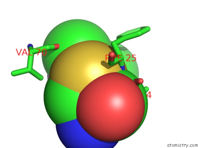
Mono view
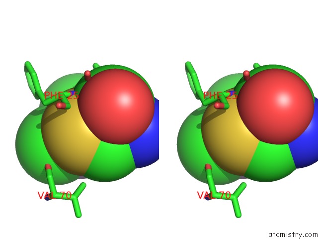
Stereo pair view
Mono view
Stereo pair view
A full contact list of Arsenic with other atoms in the As binding
site number 1 of Crystal Structures of A Poxviral Glutaredoxin in the Oxidized and Reduced States Show Redox-Correlated Structural Changes within 5.0Å range:
|
Reference:
J.P.Bacik,
B.Hazes.
Crystal Structures of A Poxviral Glutaredoxin in the Oxidized and Reduced States Show Redox-Correlated Structural Changes. J.Mol.Biol. V. 365 1545 2007.
ISSN: ISSN 0022-2836
PubMed: 17137595
DOI: 10.1016/J.JMB.2006.11.002
Page generated: Sun Jul 6 23:09:48 2025
ISSN: ISSN 0022-2836
PubMed: 17137595
DOI: 10.1016/J.JMB.2006.11.002
Last articles
F in 7JY4F in 7K0V
F in 7JYM
F in 7K15
F in 7K12
F in 7K0W
F in 7JYR
F in 7JY3
F in 7JYQ
F in 7JX9