Arsenic »
PDB 2im3-2xnq »
2ra8 »
Arsenic in PDB 2ra8: Crystal Structure of the Q64V53_BACFR Protein From Bacteroides Fragilis. Northeast Structural Genomics Consortium Target BFR43
Protein crystallography data
The structure of Crystal Structure of the Q64V53_BACFR Protein From Bacteroides Fragilis. Northeast Structural Genomics Consortium Target BFR43, PDB code: 2ra8
was solved by
S.M.Vorobiev,
M.Abashidze,
J.Seetharaman,
D.Wang,
K.Cunningham,
M.Maglaqui,
L.Owens,
R.Xiao,
T.B.Acton,
G.T.Montelione,
J.F.Hunt,
L.Tong,
Northeast Structural Genomics Consortium (Nesg),
with X-Ray Crystallography technique. A brief refinement statistics is given in the table below:
| Resolution Low / High (Å) | 19.39 / 1.95 |
| Space group | C 1 2 1 |
| Cell size a, b, c (Å), α, β, γ (°) | 108.261, 65.237, 60.008, 90.00, 111.13, 90.00 |
| R / Rfree (%) | 22.5 / 24.7 |
Arsenic Binding Sites:
The binding sites of Arsenic atom in the Crystal Structure of the Q64V53_BACFR Protein From Bacteroides Fragilis. Northeast Structural Genomics Consortium Target BFR43
(pdb code 2ra8). This binding sites where shown within
5.0 Angstroms radius around Arsenic atom.
In total only one binding site of Arsenic was determined in the Crystal Structure of the Q64V53_BACFR Protein From Bacteroides Fragilis. Northeast Structural Genomics Consortium Target BFR43, PDB code: 2ra8:
In total only one binding site of Arsenic was determined in the Crystal Structure of the Q64V53_BACFR Protein From Bacteroides Fragilis. Northeast Structural Genomics Consortium Target BFR43, PDB code: 2ra8:
Arsenic binding site 1 out of 1 in 2ra8
Go back to
Arsenic binding site 1 out
of 1 in the Crystal Structure of the Q64V53_BACFR Protein From Bacteroides Fragilis. Northeast Structural Genomics Consortium Target BFR43
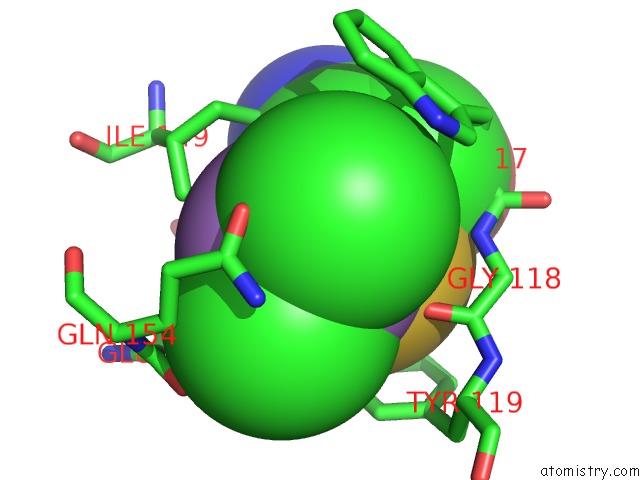
Mono view
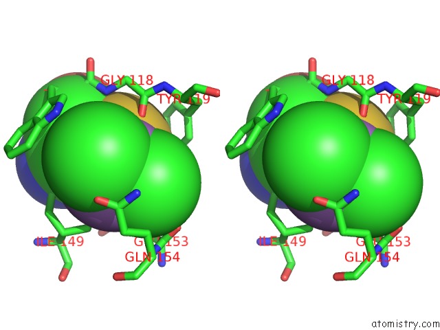
Stereo pair view
Mono view
Stereo pair view
A full contact list of Arsenic with other atoms in the As binding
site number 1 of Crystal Structure of the Q64V53_BACFR Protein From Bacteroides Fragilis. Northeast Structural Genomics Consortium Target BFR43 within 5.0Å range:
|
Reference:
S.M.Vorobiev,
M.Abashidze,
J.Seetharaman,
D.Wang,
K.Cunningham,
M.Maglaqui,
L.Owens,
R.Xiao,
T.B.Acton,
G.T.Montelione,
J.F.Hunt,
L.Tong.
Crystal Structure of the Q64V53_BACFR Protein From Bacteroides Fragilis. To Be Published.
Page generated: Sun Jul 6 23:14:01 2025
Last articles
Fe in 2YXOFe in 2YRS
Fe in 2YXC
Fe in 2YNM
Fe in 2YVJ
Fe in 2YP1
Fe in 2YU2
Fe in 2YU1
Fe in 2YQB
Fe in 2YOO