Arsenic »
PDB 2im3-2xnq »
2vdl »
Arsenic in PDB 2vdl: Re-Refinement of Integrin ALPHAIIBBETA3 Headpiece
Protein crystallography data
The structure of Re-Refinement of Integrin ALPHAIIBBETA3 Headpiece, PDB code: 2vdl
was solved by
T.A.Springer,
J.Zhu,
T.Xiao,
with X-Ray Crystallography technique. A brief refinement statistics is given in the table below:
| Resolution Low / High (Å) | 42.99 / 2.75 |
| Space group | P 32 2 1 |
| Cell size a, b, c (Å), α, β, γ (°) | 148.927, 148.927, 176.398, 90.00, 90.00, 120.00 |
| R / Rfree (%) | 14.4 / 19.1 |
Other elements in 2vdl:
The structure of Re-Refinement of Integrin ALPHAIIBBETA3 Headpiece also contains other interesting chemical elements:
| Magnesium | (Mg) | 1 atom |
| Calcium | (Ca) | 6 atoms |
Arsenic Binding Sites:
The binding sites of Arsenic atom in the Re-Refinement of Integrin ALPHAIIBBETA3 Headpiece
(pdb code 2vdl). This binding sites where shown within
5.0 Angstroms radius around Arsenic atom.
In total only one binding site of Arsenic was determined in the Re-Refinement of Integrin ALPHAIIBBETA3 Headpiece, PDB code: 2vdl:
In total only one binding site of Arsenic was determined in the Re-Refinement of Integrin ALPHAIIBBETA3 Headpiece, PDB code: 2vdl:
Arsenic binding site 1 out of 1 in 2vdl
Go back to
Arsenic binding site 1 out
of 1 in the Re-Refinement of Integrin ALPHAIIBBETA3 Headpiece
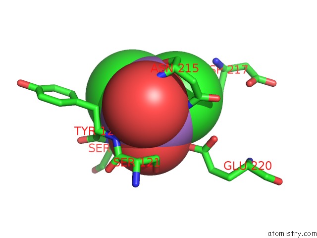
Mono view
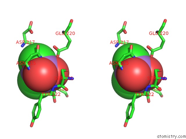
Stereo pair view
Mono view
Stereo pair view
A full contact list of Arsenic with other atoms in the As binding
site number 1 of Re-Refinement of Integrin ALPHAIIBBETA3 Headpiece within 5.0Å range:
|
Reference:
T.A.Springer,
J.Zhu,
T.Xiao.
Structural Basis For Distinctive Recognition of Fibrinogen Gammac Peptide By the Platelet Integrin ALPHAIIBBETA3. J.Cell Biol. V. 182 791 2008.
ISSN: ISSN 0021-9525
PubMed: 18710925
DOI: 10.1083/JCB.200801146
Page generated: Wed Jul 10 11:30:45 2024
ISSN: ISSN 0021-9525
PubMed: 18710925
DOI: 10.1083/JCB.200801146
Last articles
Zn in 9MJ5Zn in 9HNW
Zn in 9G0L
Zn in 9FNE
Zn in 9DZN
Zn in 9E0I
Zn in 9D32
Zn in 9DAK
Zn in 8ZXC
Zn in 8ZUF