Arsenic »
PDB 2im3-2xnq »
2vw8 »
Arsenic in PDB 2vw8: Crystal Structure of Quinolone Signal Response Protein Pqse From Pseudomonas Aeruginosa
Protein crystallography data
The structure of Crystal Structure of Quinolone Signal Response Protein Pqse From Pseudomonas Aeruginosa, PDB code: 2vw8
was solved by
L.G.Carter,
K.A.Johnson,
H.Liu,
S.A.Mcmahon,
M.Oke,
J.H.Naismith,
M.F.White,
with X-Ray Crystallography technique. A brief refinement statistics is given in the table below:
| Resolution Low / High (Å) | 51.30 / 1.45 |
| Space group | C 1 2 1 |
| Cell size a, b, c (Å), α, β, γ (°) | 103.370, 57.690, 50.240, 90.00, 97.07, 90.00 |
| R / Rfree (%) | 16.6 / 18.6 |
Other elements in 2vw8:
The structure of Crystal Structure of Quinolone Signal Response Protein Pqse From Pseudomonas Aeruginosa also contains other interesting chemical elements:
| Iron | (Fe) | 2 atoms |
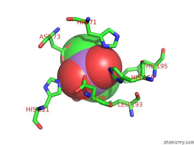
Arsenic Binding Sites:
The binding sites of Arsenic atom in the Crystal Structure of Quinolone Signal Response Protein Pqse From Pseudomonas Aeruginosa
(pdb code 2vw8). This binding sites where shown within
5.0 Angstroms radius around Arsenic atom.
In total only one binding site of Arsenic was determined in the Crystal Structure of Quinolone Signal Response Protein Pqse From Pseudomonas Aeruginosa, PDB code: 2vw8:
In total only one binding site of Arsenic was determined in the Crystal Structure of Quinolone Signal Response Protein Pqse From Pseudomonas Aeruginosa, PDB code: 2vw8:
Arsenic binding site 1 out of 1 in 2vw8
Go back to
Arsenic binding site 1 out
of 1 in the Crystal Structure of Quinolone Signal Response Protein Pqse From Pseudomonas Aeruginosa
Mono view
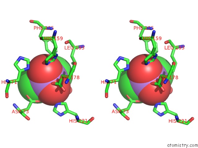
Stereo pair view
Mono view
Stereo pair view
A full contact list of Arsenic with other atoms in the As binding
site number 1 of Crystal Structure of Quinolone Signal Response Protein Pqse From Pseudomonas Aeruginosa within 5.0Å range:
|
Reference:
M.Oke,
L.G.Carter,
K.A.Johnson,
H.Liu,
S.A.Mcmahon,
X.Yan,
M.Kerou,
N.D.Weikart,
N.Kadi,
M.A.Sheikh,
S.Schmelz,
M.Dorward,
M.Zawadzki,
C.Cozens,
H.Falconer,
H.Powers,
I.M.Overton,
C.A.J.Van Niekerk,
X.Peng,
P.Patel,
R.A.Garrett,
D.Prangishvili C,
H.Botting,
P.J.Coote,
D.T.F.Dryden,
G.J.Barton,
U.Schwarz-Linek,
G.L.Challis,
G.L.Taylor,
M.F.White,
J.H.Naismith.
The Scottish Structural Proteomics Facility: Targets, Methods and Outputs. J.Struct.Funct.Genomics V. 11 167 2010.
ISSN: ISSN 1345-711X
PubMed: 20419351
DOI: 10.1007/S10969-010-9090-Y
Page generated: Sun Jul 6 23:16:44 2025
ISSN: ISSN 1345-711X
PubMed: 20419351
DOI: 10.1007/S10969-010-9090-Y
Last articles
Cl in 5V6SCl in 5V5Z
Cl in 5V69
Cl in 5V4H
Cl in 5V53
Cl in 5V4I
Cl in 5V4Q
Cl in 5V36
Cl in 5V4G
Cl in 5V2C