Arsenic »
PDB 2xod-3g2f »
3bft »
Arsenic in PDB 3bft: Structure of the Ligand-Binding Core of GLUR2 in Complex with the Agonist (S)-Tdpa at 2.25 A Resolution
Protein crystallography data
The structure of Structure of the Ligand-Binding Core of GLUR2 in Complex with the Agonist (S)-Tdpa at 2.25 A Resolution, PDB code: 3bft
was solved by
M.Beich-Frandsen,
O.Mirza,
B.Vestergaard,
M.Gajhede,
J.S.Kastrup,
with X-Ray Crystallography technique. A brief refinement statistics is given in the table below:
| Resolution Low / High (Å) | 20.00 / 2.27 |
| Space group | P 21 21 2 |
| Cell size a, b, c (Å), α, β, γ (°) | 113.820, 163.333, 47.270, 90.00, 90.00, 90.00 |
| R / Rfree (%) | 20 / 25 |
Other elements in 3bft:
The structure of Structure of the Ligand-Binding Core of GLUR2 in Complex with the Agonist (S)-Tdpa at 2.25 A Resolution also contains other interesting chemical elements:
| Zinc | (Zn) | 5 atoms |
| Chlorine | (Cl) | 1 atom |
| Sodium | (Na) | 2 atoms |
Arsenic Binding Sites:
The binding sites of Arsenic atom in the Structure of the Ligand-Binding Core of GLUR2 in Complex with the Agonist (S)-Tdpa at 2.25 A Resolution
(pdb code 3bft). This binding sites where shown within
5.0 Angstroms radius around Arsenic atom.
In total only one binding site of Arsenic was determined in the Structure of the Ligand-Binding Core of GLUR2 in Complex with the Agonist (S)-Tdpa at 2.25 A Resolution, PDB code: 3bft:
In total only one binding site of Arsenic was determined in the Structure of the Ligand-Binding Core of GLUR2 in Complex with the Agonist (S)-Tdpa at 2.25 A Resolution, PDB code: 3bft:
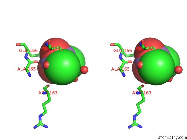
Arsenic binding site 1 out of 1 in 3bft
Go back to
Arsenic binding site 1 out
of 1 in the Structure of the Ligand-Binding Core of GLUR2 in Complex with the Agonist (S)-Tdpa at 2.25 A Resolution
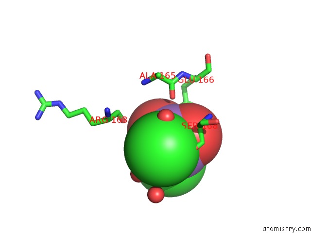
Mono view
Stereo pair view
Mono view
Stereo pair view
A full contact list of Arsenic with other atoms in the As binding
site number 1 of Structure of the Ligand-Binding Core of GLUR2 in Complex with the Agonist (S)-Tdpa at 2.25 A Resolution within 5.0Å range:
|
Reference:
M.Beich-Frandsen,
D.S.Pickering,
O.Mirza,
T.N.Johansen,
J.Greenwood,
B.Vestergaard,
A.Schousboe,
M.Gajhede,
T.Liljefors,
J.S.Kastrup.
Structures of the Ligand-Binding Core of IGLUR2 in Complex with the Agonists (R)- and (S)-2-Amino-3-(4-Hydroxy-1,2,5-Thiadiazol-3-Yl)Propionic Acid Explain Their Unusual Equipotency. J.Med.Chem. V. 51 1459 2008.
ISSN: ISSN 0022-2623
PubMed: 18269227
DOI: 10.1021/JM701126W
Page generated: Sun Jul 6 23:19:20 2025
ISSN: ISSN 0022-2623
PubMed: 18269227
DOI: 10.1021/JM701126W
Last articles
F in 4D7OF in 4D44
F in 4D6Y
F in 4D7J
F in 4D7I
F in 4D7H
F in 4D6U
F in 4D6T
F in 4D5H
F in 4D42