Arsenic »
PDB 2xod-3g2f »
3e3z »
Arsenic in PDB 3e3z: Crystal Structure of Bovine Coupling Factor B Bound with Phenylarsine Oxide
Enzymatic activity of Crystal Structure of Bovine Coupling Factor B Bound with Phenylarsine Oxide
All present enzymatic activity of Crystal Structure of Bovine Coupling Factor B Bound with Phenylarsine Oxide:
3.6.3.14;
3.6.3.14;
Protein crystallography data
The structure of Crystal Structure of Bovine Coupling Factor B Bound with Phenylarsine Oxide, PDB code: 3e3z
was solved by
R.M.Stroud,
J.K.Lee,
G.I.Belogrudov,
with X-Ray Crystallography technique. A brief refinement statistics is given in the table below:
| Resolution Low / High (Å) | 30.00 / 1.70 |
| Space group | C 1 2 1 |
| Cell size a, b, c (Å), α, β, γ (°) | 106.122, 49.722, 36.902, 90.00, 108.84, 90.00 |
| R / Rfree (%) | 14.9 / 18.8 |
Other elements in 3e3z:
The structure of Crystal Structure of Bovine Coupling Factor B Bound with Phenylarsine Oxide also contains other interesting chemical elements:
| Magnesium | (Mg) | 1 atom |
Arsenic Binding Sites:
The binding sites of Arsenic atom in the Crystal Structure of Bovine Coupling Factor B Bound with Phenylarsine Oxide
(pdb code 3e3z). This binding sites where shown within
5.0 Angstroms radius around Arsenic atom.
In total only one binding site of Arsenic was determined in the Crystal Structure of Bovine Coupling Factor B Bound with Phenylarsine Oxide, PDB code: 3e3z:
In total only one binding site of Arsenic was determined in the Crystal Structure of Bovine Coupling Factor B Bound with Phenylarsine Oxide, PDB code: 3e3z:
Arsenic binding site 1 out of 1 in 3e3z
Go back to
Arsenic binding site 1 out
of 1 in the Crystal Structure of Bovine Coupling Factor B Bound with Phenylarsine Oxide
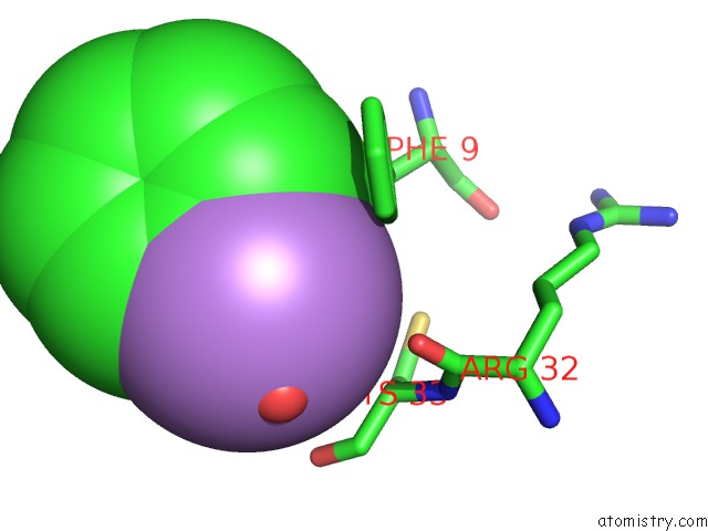
Mono view
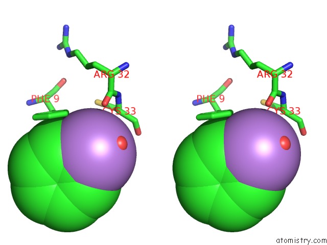
Stereo pair view
Mono view
Stereo pair view
A full contact list of Arsenic with other atoms in the As binding
site number 1 of Crystal Structure of Bovine Coupling Factor B Bound with Phenylarsine Oxide within 5.0Å range:
|
Reference:
J.K.Lee,
G.I.Belogrudov,
R.M.Stroud.
Crystal Structure of Bovine Mitochondrial Factor B at 0.96-A Resolution. Proc.Natl.Acad.Sci.Usa V. 105 13379 2008.
ISSN: ISSN 0027-8424
PubMed: 18768789
DOI: 10.1073/PNAS.0805689105
Page generated: Sun Jul 6 23:21:06 2025
ISSN: ISSN 0027-8424
PubMed: 18768789
DOI: 10.1073/PNAS.0805689105
Last articles
Cl in 8AM2Cl in 8AM4
Cl in 8ALW
Cl in 8AM0
Cl in 8ALV
Cl in 8ALT
Cl in 8AEN
Cl in 8AKG
Cl in 8ALN
Cl in 8ALM