Arsenic »
PDB 2xod-3g2f »
3fmu »
Arsenic in PDB 3fmu: Crystal Structure Analysis of Fungal Versatile Peroxidase From Pleurotus Eryngii
Enzymatic activity of Crystal Structure Analysis of Fungal Versatile Peroxidase From Pleurotus Eryngii
All present enzymatic activity of Crystal Structure Analysis of Fungal Versatile Peroxidase From Pleurotus Eryngii:
1.11.1.16;
1.11.1.16;
Protein crystallography data
The structure of Crystal Structure Analysis of Fungal Versatile Peroxidase From Pleurotus Eryngii, PDB code: 3fmu
was solved by
K.Piontek,
A.T.Martinez,
T.Choinowski,
D.A.Plattner,
with X-Ray Crystallography technique. A brief refinement statistics is given in the table below:
| Resolution Low / High (Å) | 44.69 / 1.04 |
| Space group | P 43 |
| Cell size a, b, c (Å), α, β, γ (°) | 63.199, 63.199, 99.237, 90.00, 90.00, 90.00 |
| R / Rfree (%) | 11.1 / 12.3 |
Other elements in 3fmu:
The structure of Crystal Structure Analysis of Fungal Versatile Peroxidase From Pleurotus Eryngii also contains other interesting chemical elements:
| Zinc | (Zn) | 8 atoms |
| Iron | (Fe) | 6 atoms |
| Calcium | (Ca) | 2 atoms |
Arsenic Binding Sites:
The binding sites of Arsenic atom in the Crystal Structure Analysis of Fungal Versatile Peroxidase From Pleurotus Eryngii
(pdb code 3fmu). This binding sites where shown within
5.0 Angstroms radius around Arsenic atom.
In total only one binding site of Arsenic was determined in the Crystal Structure Analysis of Fungal Versatile Peroxidase From Pleurotus Eryngii, PDB code: 3fmu:
In total only one binding site of Arsenic was determined in the Crystal Structure Analysis of Fungal Versatile Peroxidase From Pleurotus Eryngii, PDB code: 3fmu:
Arsenic binding site 1 out of 1 in 3fmu
Go back to
Arsenic binding site 1 out
of 1 in the Crystal Structure Analysis of Fungal Versatile Peroxidase From Pleurotus Eryngii

Mono view
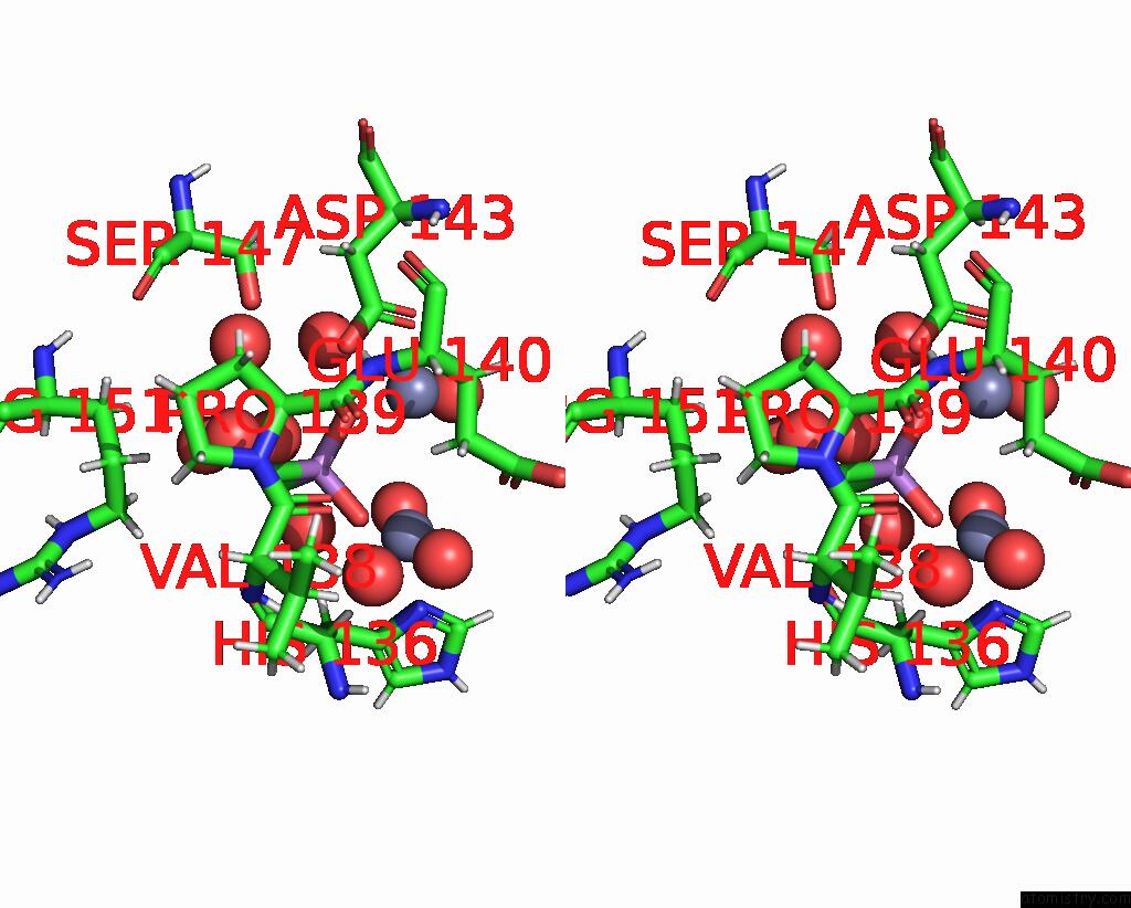
Stereo pair view
Mono view
Stereo pair view
A full contact list of Arsenic with other atoms in the As binding
site number 1 of Crystal Structure Analysis of Fungal Versatile Peroxidase From Pleurotus Eryngii within 5.0Å range:
|
Reference:
K.Piontek,
T.Choinowski,
M.Perez-Boada,
F.J.Ruiz-Duenas,
M.J.Martinez,
D.A.Plattner,
A.T.Martinez.
Structural and Site-Directed Mutagenesis Study of Versatile Peroxidase Oxidizing Both Mn(II) and Aromatic Substrates To Be Published.
Page generated: Wed Jul 10 11:40:06 2024
Last articles
Zn in 9MJ5Zn in 9HNW
Zn in 9G0L
Zn in 9FNE
Zn in 9DZN
Zn in 9E0I
Zn in 9D32
Zn in 9DAK
Zn in 8ZXC
Zn in 8ZUF