Arsenic »
PDB 3g3s-3n5t »
3h6n »
Arsenic in PDB 3h6n: Crystal Structure of the Ubiquitin-Like Domain of Plexin D1
Protein crystallography data
The structure of Crystal Structure of the Ubiquitin-Like Domain of Plexin D1, PDB code: 3h6n
was solved by
Y.Tong,
L.Nedyalkova,
W.Tempel,
F.Mackenzie,
C.H.Arrowsmith,
A.M.Edwards,
C.Bountra,
J.Weigelt,
A.Bochkarev,
M.Buck,
H.Park,
Structural Genomicsconsortium (Sgc),
with X-Ray Crystallography technique. A brief refinement statistics is given in the table below:
| Resolution Low / High (Å) | 30.00 / 2.00 |
| Space group | C 1 2 1 |
| Cell size a, b, c (Å), α, β, γ (°) | 81.777, 27.051, 52.653, 90.00, 114.06, 90.00 |
| R / Rfree (%) | 24.3 / 27.6 |
Arsenic Binding Sites:
The binding sites of Arsenic atom in the Crystal Structure of the Ubiquitin-Like Domain of Plexin D1
(pdb code 3h6n). This binding sites where shown within
5.0 Angstroms radius around Arsenic atom.
In total 2 binding sites of Arsenic where determined in the Crystal Structure of the Ubiquitin-Like Domain of Plexin D1, PDB code: 3h6n:
Jump to Arsenic binding site number: 1; 2;
In total 2 binding sites of Arsenic where determined in the Crystal Structure of the Ubiquitin-Like Domain of Plexin D1, PDB code: 3h6n:
Jump to Arsenic binding site number: 1; 2;
Arsenic binding site 1 out of 2 in 3h6n
Go back to
Arsenic binding site 1 out
of 2 in the Crystal Structure of the Ubiquitin-Like Domain of Plexin D1
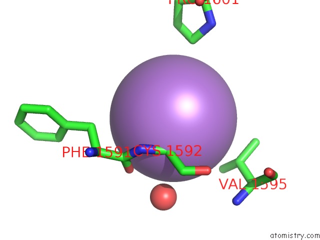
Mono view
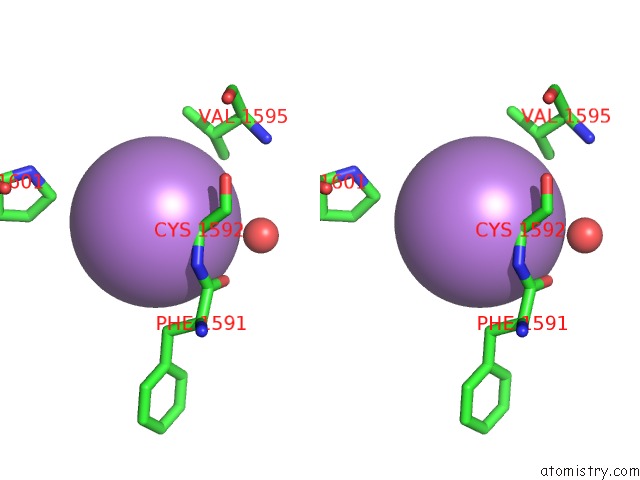
Stereo pair view
Mono view
Stereo pair view
A full contact list of Arsenic with other atoms in the As binding
site number 1 of Crystal Structure of the Ubiquitin-Like Domain of Plexin D1 within 5.0Å range:
|
Arsenic binding site 2 out of 2 in 3h6n
Go back to
Arsenic binding site 2 out
of 2 in the Crystal Structure of the Ubiquitin-Like Domain of Plexin D1
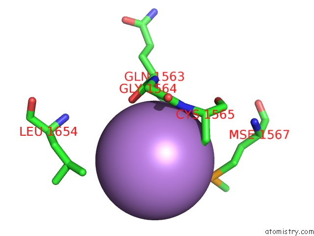
Mono view
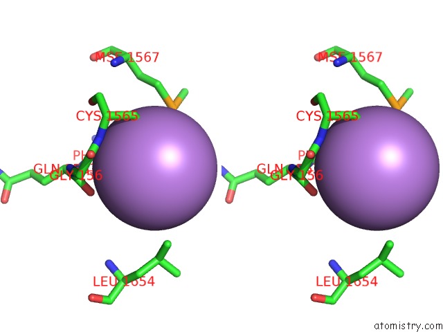
Stereo pair view
Mono view
Stereo pair view
A full contact list of Arsenic with other atoms in the As binding
site number 2 of Crystal Structure of the Ubiquitin-Like Domain of Plexin D1 within 5.0Å range:
|
Reference:
Y.Tong,
L.Nedyalkova,
W.Tempel,
F.Mackenzie,
C.H.Arrowsmith,
A.M.Edwards,
C.Bountra,
J.Weigelt,
A.Bochkarev,
M.Buck,
H.Park.
Crystal Structure of the Ubiquitin-Like Domain of Plexin D1 To Be Published.
Page generated: Sun Jul 6 23:24:02 2025
Last articles
Cl in 8AY6Cl in 8AXT
Cl in 8AXS
Cl in 8AXW
Cl in 8AXU
Cl in 8AXI
Cl in 8AXC
Cl in 8AXR
Cl in 8AWW
Cl in 8AXE