Arsenic »
PDB 3g3s-3n5t »
3h6t »
Arsenic in PDB 3h6t: Crystal Structure of the IGLUR2 Ligand-Binding Core (S1S2J-N754S) in Complex with Glutamate and Cyclothiazide at 2.25 A Resolution
Protein crystallography data
The structure of Crystal Structure of the IGLUR2 Ligand-Binding Core (S1S2J-N754S) in Complex with Glutamate and Cyclothiazide at 2.25 A Resolution, PDB code: 3h6t
was solved by
H.Hald,
M.Gajhede,
J.S.Kastrup,
with X-Ray Crystallography technique. A brief refinement statistics is given in the table below:
| Resolution Low / High (Å) | 22.84 / 2.25 |
| Space group | P 21 21 2 |
| Cell size a, b, c (Å), α, β, γ (°) | 114.486, 163.136, 47.354, 90.00, 90.00, 90.00 |
| R / Rfree (%) | 18.2 / 22.8 |
Other elements in 3h6t:
The structure of Crystal Structure of the IGLUR2 Ligand-Binding Core (S1S2J-N754S) in Complex with Glutamate and Cyclothiazide at 2.25 A Resolution also contains other interesting chemical elements:
| Chlorine | (Cl) | 3 atoms |
| Zinc | (Zn) | 10 atoms |
Arsenic Binding Sites:
The binding sites of Arsenic atom in the Crystal Structure of the IGLUR2 Ligand-Binding Core (S1S2J-N754S) in Complex with Glutamate and Cyclothiazide at 2.25 A Resolution
(pdb code 3h6t). This binding sites where shown within
5.0 Angstroms radius around Arsenic atom.
In total only one binding site of Arsenic was determined in the Crystal Structure of the IGLUR2 Ligand-Binding Core (S1S2J-N754S) in Complex with Glutamate and Cyclothiazide at 2.25 A Resolution, PDB code: 3h6t:
In total only one binding site of Arsenic was determined in the Crystal Structure of the IGLUR2 Ligand-Binding Core (S1S2J-N754S) in Complex with Glutamate and Cyclothiazide at 2.25 A Resolution, PDB code: 3h6t:
Arsenic binding site 1 out of 1 in 3h6t
Go back to
Arsenic binding site 1 out
of 1 in the Crystal Structure of the IGLUR2 Ligand-Binding Core (S1S2J-N754S) in Complex with Glutamate and Cyclothiazide at 2.25 A Resolution
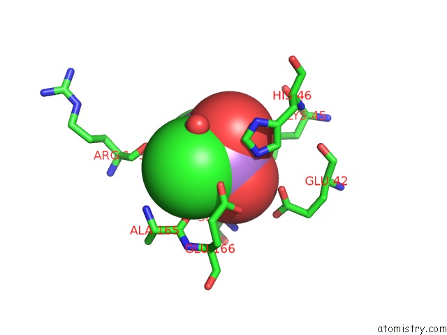
Mono view
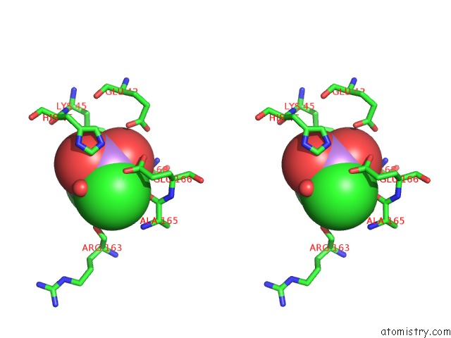
Stereo pair view
Mono view
Stereo pair view
A full contact list of Arsenic with other atoms in the As binding
site number 1 of Crystal Structure of the IGLUR2 Ligand-Binding Core (S1S2J-N754S) in Complex with Glutamate and Cyclothiazide at 2.25 A Resolution within 5.0Å range:
|
Reference:
H.Hald,
P.K.Ahring,
D.B.Timmermann,
T.Liljefors,
M.Gajhede,
J.S.Kastrup.
Distinct Structural Features of Cyclothiazide Are Responsible For Effects on Peak Current Amplitude and Desensitization Kinetics at IGLUR2. J.Mol.Biol. V. 391 906 2009.
ISSN: ISSN 0022-2836
PubMed: 19591837
DOI: 10.1016/J.JMB.2009.07.002
Page generated: Wed Jul 10 11:43:02 2024
ISSN: ISSN 0022-2836
PubMed: 19591837
DOI: 10.1016/J.JMB.2009.07.002
Last articles
Zn in 9MJ5Zn in 9HNW
Zn in 9G0L
Zn in 9FNE
Zn in 9DZN
Zn in 9E0I
Zn in 9D32
Zn in 9DAK
Zn in 8ZXC
Zn in 8ZUF