Arsenic »
PDB 3n67-3smt »
3pb6 »
Arsenic in PDB 3pb6: Crystal Structure of the Catalytic Domain of Human Golgi-Resident Glutaminyl Cyclase at pH 6.5
Enzymatic activity of Crystal Structure of the Catalytic Domain of Human Golgi-Resident Glutaminyl Cyclase at pH 6.5
All present enzymatic activity of Crystal Structure of the Catalytic Domain of Human Golgi-Resident Glutaminyl Cyclase at pH 6.5:
2.3.2.5;
2.3.2.5;
Protein crystallography data
The structure of Crystal Structure of the Catalytic Domain of Human Golgi-Resident Glutaminyl Cyclase at pH 6.5, PDB code: 3pb6
was solved by
K.F.Huang,
S.S.Liaw,
W.L.Huang,
C.Y.Chia,
Y.C.Lo,
Y.L.Chen,
A.H.J.Wang,
with X-Ray Crystallography technique. A brief refinement statistics is given in the table below:
| Resolution Low / High (Å) | 30.00 / 1.05 |
| Space group | P 21 21 21 |
| Cell size a, b, c (Å), α, β, γ (°) | 53.097, 68.527, 77.348, 90.00, 90.00, 90.00 |
| R / Rfree (%) | 13.8 / 15.8 |
Other elements in 3pb6:
The structure of Crystal Structure of the Catalytic Domain of Human Golgi-Resident Glutaminyl Cyclase at pH 6.5 also contains other interesting chemical elements:
| Zinc | (Zn) | 1 atom |
Arsenic Binding Sites:
The binding sites of Arsenic atom in the Crystal Structure of the Catalytic Domain of Human Golgi-Resident Glutaminyl Cyclase at pH 6.5
(pdb code 3pb6). This binding sites where shown within
5.0 Angstroms radius around Arsenic atom.
In total only one binding site of Arsenic was determined in the Crystal Structure of the Catalytic Domain of Human Golgi-Resident Glutaminyl Cyclase at pH 6.5, PDB code: 3pb6:
In total only one binding site of Arsenic was determined in the Crystal Structure of the Catalytic Domain of Human Golgi-Resident Glutaminyl Cyclase at pH 6.5, PDB code: 3pb6:
Arsenic binding site 1 out of 1 in 3pb6
Go back to
Arsenic binding site 1 out
of 1 in the Crystal Structure of the Catalytic Domain of Human Golgi-Resident Glutaminyl Cyclase at pH 6.5
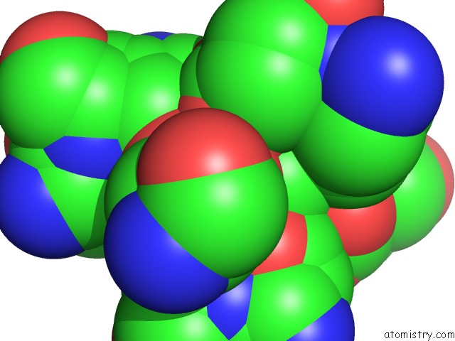
Mono view
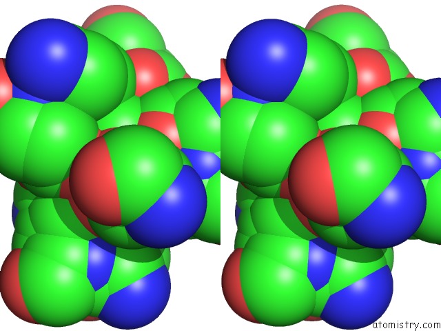
Stereo pair view
Mono view
Stereo pair view
A full contact list of Arsenic with other atoms in the As binding
site number 1 of Crystal Structure of the Catalytic Domain of Human Golgi-Resident Glutaminyl Cyclase at pH 6.5 within 5.0Å range:
|
Reference:
K.F.Huang,
S.S.Liaw,
W.L.Huang,
C.Y.Chia,
Y.C.Lo,
Y.L.Chen,
A.H.J.Wang.
Structures of Human Golgi-Resident Glutaminyl Cyclase and Its Complexes with Inhibitors Reveal A Large Loop Movement Upon Inhibitor Binding J.Biol.Chem. V. 286 12439 2011.
ISSN: ISSN 0021-9258
PubMed: 21288892
DOI: 10.1074/JBC.M110.208595
Page generated: Sun Jul 6 23:31:27 2025
ISSN: ISSN 0021-9258
PubMed: 21288892
DOI: 10.1074/JBC.M110.208595
Last articles
Cl in 2WJ2Cl in 2WJ9
Cl in 2WJ1
Cl in 2WIK
Cl in 2WIL
Cl in 2WIG
Cl in 2WIJ
Cl in 2WID
Cl in 2WIF
Cl in 2WHI