Arsenic »
PDB 3n67-3smt »
3pqs »
Arsenic in PDB 3pqs: The Crystal Structures of Porcine Pathogen APH87_TBPB
Protein crystallography data
The structure of The Crystal Structures of Porcine Pathogen APH87_TBPB, PDB code: 3pqs
was solved by
C.Calmettes,
T.F.Moraes,
with X-Ray Crystallography technique. A brief refinement statistics is given in the table below:
| Resolution Low / High (Å) | 47.14 / 2.10 |
| Space group | C 2 2 21 |
| Cell size a, b, c (Å), α, β, γ (°) | 132.600, 151.300, 90.270, 90.00, 90.00, 90.00 |
| R / Rfree (%) | 16 / 19.7 |
Arsenic Binding Sites:
The binding sites of Arsenic atom in the The Crystal Structures of Porcine Pathogen APH87_TBPB
(pdb code 3pqs). This binding sites where shown within
5.0 Angstroms radius around Arsenic atom.
In total only one binding site of Arsenic was determined in the The Crystal Structures of Porcine Pathogen APH87_TBPB, PDB code: 3pqs:
In total only one binding site of Arsenic was determined in the The Crystal Structures of Porcine Pathogen APH87_TBPB, PDB code: 3pqs:
Arsenic binding site 1 out of 1 in 3pqs
Go back to
Arsenic binding site 1 out
of 1 in the The Crystal Structures of Porcine Pathogen APH87_TBPB
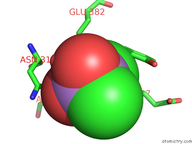
Mono view
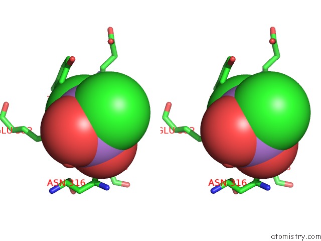
Stereo pair view
Mono view
Stereo pair view
A full contact list of Arsenic with other atoms in the As binding
site number 1 of The Crystal Structures of Porcine Pathogen APH87_TBPB within 5.0Å range:
|
Reference:
C.Calmettes,
R.H.Yu,
L.P.Silva,
D.Curran,
D.C.Schriemer,
A.B.Schryvers,
T.F.Moraes.
Structural Variations Within the Transferrin Binding Site on Transferrin-Binding Protein B, Tbpb. J.Biol.Chem. V. 286 12683 2011.
ISSN: ISSN 0021-9258
PubMed: 21297163
DOI: 10.1074/JBC.M110.206102
Page generated: Sun Jul 6 23:31:50 2025
ISSN: ISSN 0021-9258
PubMed: 21297163
DOI: 10.1074/JBC.M110.206102
Last articles
Fe in 2YXOFe in 2YRS
Fe in 2YXC
Fe in 2YNM
Fe in 2YVJ
Fe in 2YP1
Fe in 2YU2
Fe in 2YU1
Fe in 2YQB
Fe in 2YOO