Arsenic »
PDB 3n67-3smt »
3rhg »
Arsenic in PDB 3rhg: Crystal Structure of Amidohydrolase PMI1525 (Target Efi-500319) From Proteus Mirabilis HI4320
Protein crystallography data
The structure of Crystal Structure of Amidohydrolase PMI1525 (Target Efi-500319) From Proteus Mirabilis HI4320, PDB code: 3rhg
was solved by
Y.Patskovsky,
B.Hillerich,
R.D.Seidel,
W.D.Zencheck,
R.Toro,
H.J.Imker,
F.M.Raushel,
J.A.Gerlt,
S.C.Almo,
Enzyme Function Initiative (Efi),
with X-Ray Crystallography technique. A brief refinement statistics is given in the table below:
| Resolution Low / High (Å) | 50.00 / 1.53 |
| Space group | P 32 2 1 |
| Cell size a, b, c (Å), α, β, γ (°) | 101.203, 101.203, 65.614, 90.00, 90.00, 120.00 |
| R / Rfree (%) | 12 / 15.6 |
Other elements in 3rhg:
The structure of Crystal Structure of Amidohydrolase PMI1525 (Target Efi-500319) From Proteus Mirabilis HI4320 also contains other interesting chemical elements:
| Zinc | (Zn) | 2 atoms |
Arsenic Binding Sites:
The binding sites of Arsenic atom in the Crystal Structure of Amidohydrolase PMI1525 (Target Efi-500319) From Proteus Mirabilis HI4320
(pdb code 3rhg). This binding sites where shown within
5.0 Angstroms radius around Arsenic atom.
In total only one binding site of Arsenic was determined in the Crystal Structure of Amidohydrolase PMI1525 (Target Efi-500319) From Proteus Mirabilis HI4320, PDB code: 3rhg:
In total only one binding site of Arsenic was determined in the Crystal Structure of Amidohydrolase PMI1525 (Target Efi-500319) From Proteus Mirabilis HI4320, PDB code: 3rhg:
Arsenic binding site 1 out of 1 in 3rhg
Go back to
Arsenic binding site 1 out
of 1 in the Crystal Structure of Amidohydrolase PMI1525 (Target Efi-500319) From Proteus Mirabilis HI4320
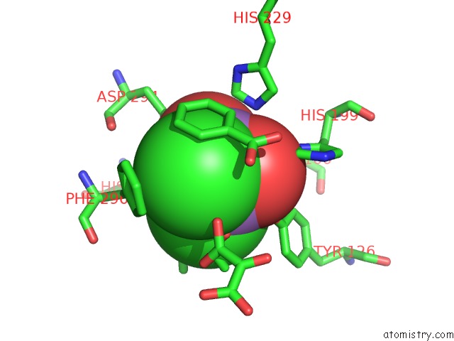
Mono view
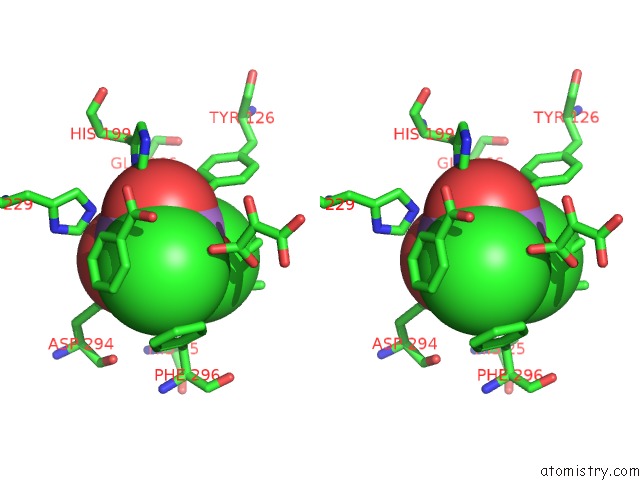
Stereo pair view
Mono view
Stereo pair view
A full contact list of Arsenic with other atoms in the As binding
site number 1 of Crystal Structure of Amidohydrolase PMI1525 (Target Efi-500319) From Proteus Mirabilis HI4320 within 5.0Å range:
|
Reference:
Y.Patskovsky,
B.Hillerich,
R.D.Seidel,
W.D.Zencheck,
R.Toro,
H.J.Imker,
F.M.Raushel,
J.A.Gerlt,
S.C.Almo.
Crystal Structure of Amidohydrolase PMI1525 From Proteus Mirabilis HI4320 To Be Published.
Page generated: Sun Jul 6 23:32:21 2025
Last articles
Cl in 2QG4Cl in 2QHF
Cl in 2QGW
Cl in 2QF7
Cl in 2QA4
Cl in 2QEI
Cl in 2QEP
Cl in 2QE5
Cl in 2QE3
Cl in 2QDY