Arsenic »
PDB 4mwd-4uha »
4pcp »
Arsenic in PDB 4pcp: Crystal Structure of Phosphotriesterase Variant R0
Enzymatic activity of Crystal Structure of Phosphotriesterase Variant R0
All present enzymatic activity of Crystal Structure of Phosphotriesterase Variant R0:
3.1.8.1;
3.1.8.1;
Protein crystallography data
The structure of Crystal Structure of Phosphotriesterase Variant R0, PDB code: 4pcp
was solved by
E.Campbell,
M.Kaltenbach,
N.Tokuriki,
C.J.Jackson,
with X-Ray Crystallography technique. A brief refinement statistics is given in the table below:
| Resolution Low / High (Å) | 43.16 / 1.63 |
| Space group | P 21 21 2 |
| Cell size a, b, c (Å), α, β, γ (°) | 86.141, 86.325, 88.722, 90.00, 90.00, 90.00 |
| R / Rfree (%) | 17.9 / 21.9 |
Other elements in 4pcp:
The structure of Crystal Structure of Phosphotriesterase Variant R0 also contains other interesting chemical elements:
| Zinc | (Zn) | 4 atoms |
Arsenic Binding Sites:
The binding sites of Arsenic atom in the Crystal Structure of Phosphotriesterase Variant R0
(pdb code 4pcp). This binding sites where shown within
5.0 Angstroms radius around Arsenic atom.
In total 2 binding sites of Arsenic where determined in the Crystal Structure of Phosphotriesterase Variant R0, PDB code: 4pcp:
Jump to Arsenic binding site number: 1; 2;
In total 2 binding sites of Arsenic where determined in the Crystal Structure of Phosphotriesterase Variant R0, PDB code: 4pcp:
Jump to Arsenic binding site number: 1; 2;
Arsenic binding site 1 out of 2 in 4pcp
Go back to
Arsenic binding site 1 out
of 2 in the Crystal Structure of Phosphotriesterase Variant R0
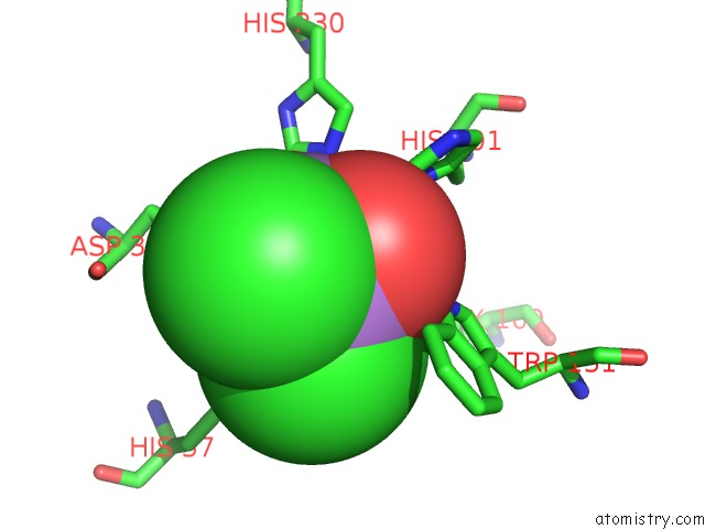
Mono view

Stereo pair view
Mono view
Stereo pair view
A full contact list of Arsenic with other atoms in the As binding
site number 1 of Crystal Structure of Phosphotriesterase Variant R0 within 5.0Å range:
|
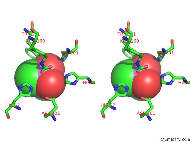
Arsenic binding site 2 out of 2 in 4pcp
Go back to
Arsenic binding site 2 out
of 2 in the Crystal Structure of Phosphotriesterase Variant R0

Mono view
Stereo pair view
Mono view
Stereo pair view
A full contact list of Arsenic with other atoms in the As binding
site number 2 of Crystal Structure of Phosphotriesterase Variant R0 within 5.0Å range:
|
Reference:
E.Campbell,
M.Kaltenbach,
N.Tokuriki,
C.Jackson.
Crystal Structure of Phosphotriesterase Variant R0 To Be Published.
Page generated: Sun Jul 6 23:57:34 2025
Last articles
F in 7LDEF in 7LEP
F in 7LDD
F in 7LGX
F in 7LGK
F in 7LG8
F in 7LD3
F in 7LCR
F in 7LCM
F in 7LCO