Arsenic »
PDB 4mwd-4uha »
4u2v »
Arsenic in PDB 4u2v: Bak BH3-in-Groove Dimer (Gfp)
Protein crystallography data
The structure of Bak BH3-in-Groove Dimer (Gfp), PDB code: 4u2v
was solved by
J.M.Brouwer,
P.M.Colman,
P.E.Czabotar,
with X-Ray Crystallography technique. A brief refinement statistics is given in the table below:
| Resolution Low / High (Å) | 20.00 / 2.30 |
| Space group | P 1 21 1 |
| Cell size a, b, c (Å), α, β, γ (°) | 109.190, 63.840, 122.020, 90.00, 98.35, 90.00 |
| R / Rfree (%) | 19.8 / 25 |
Arsenic Binding Sites:
The binding sites of Arsenic atom in the Bak BH3-in-Groove Dimer (Gfp)
(pdb code 4u2v). This binding sites where shown within
5.0 Angstroms radius around Arsenic atom.
In total 6 binding sites of Arsenic where determined in the Bak BH3-in-Groove Dimer (Gfp), PDB code: 4u2v:
Jump to Arsenic binding site number: 1; 2; 3; 4; 5; 6;
In total 6 binding sites of Arsenic where determined in the Bak BH3-in-Groove Dimer (Gfp), PDB code: 4u2v:
Jump to Arsenic binding site number: 1; 2; 3; 4; 5; 6;
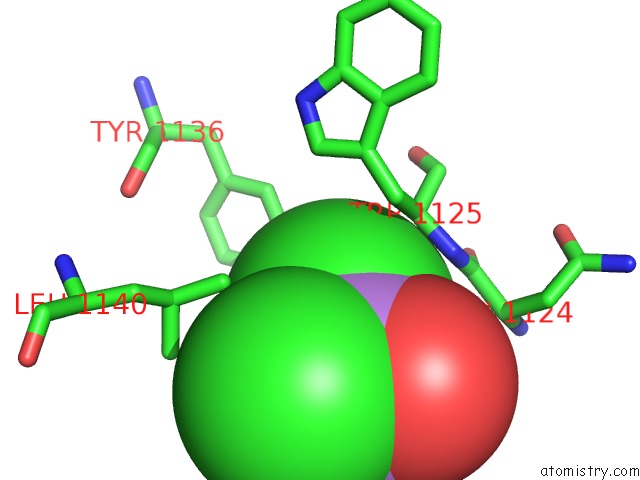
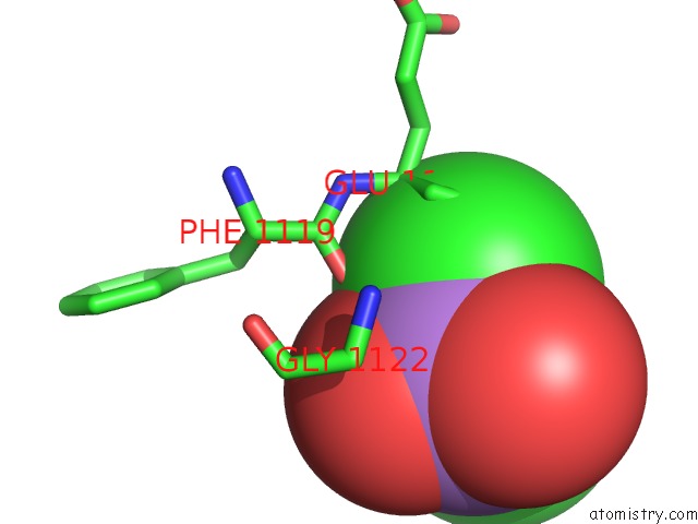
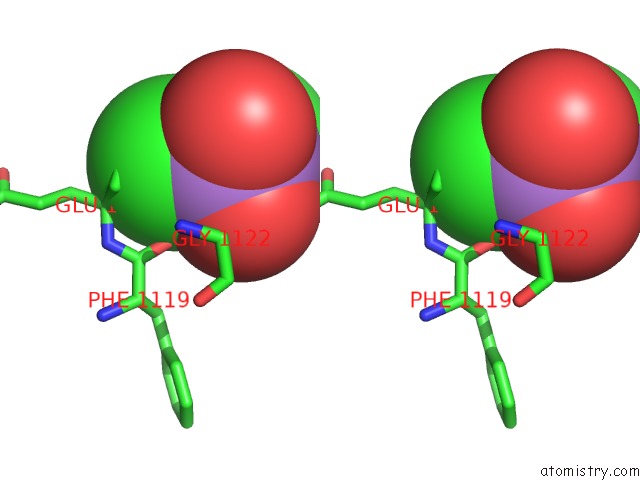
Arsenic binding site 1 out of 6 in 4u2v
Go back to
Arsenic binding site 1 out
of 6 in the Bak BH3-in-Groove Dimer (Gfp)
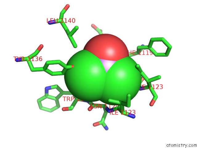
Mono view
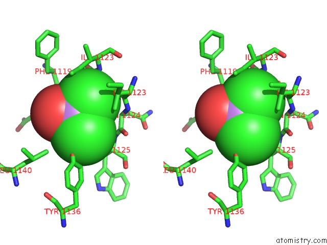
Stereo pair view
Mono view
Stereo pair view
A full contact list of Arsenic with other atoms in the As binding
site number 1 of Bak BH3-in-Groove Dimer (Gfp) within 5.0Å range:
|
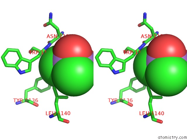
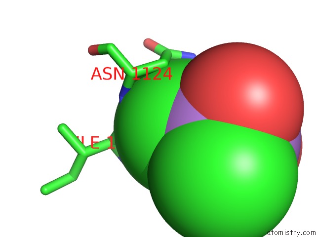
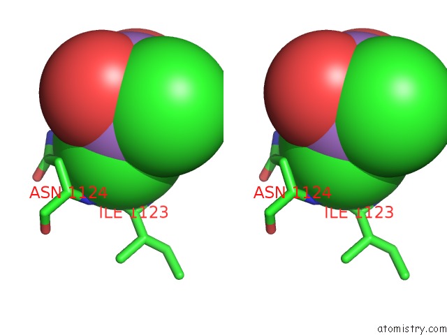
Arsenic binding site 2 out of 6 in 4u2v
Go back to
Arsenic binding site 2 out
of 6 in the Bak BH3-in-Groove Dimer (Gfp)
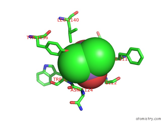
Mono view
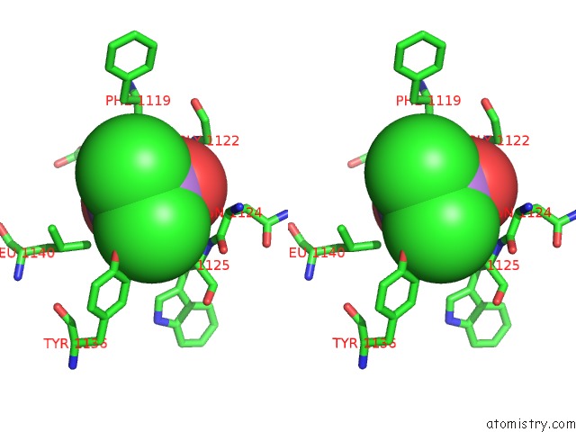
Stereo pair view
Mono view
Stereo pair view
A full contact list of Arsenic with other atoms in the As binding
site number 2 of Bak BH3-in-Groove Dimer (Gfp) within 5.0Å range:
|
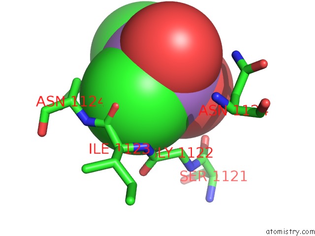
Arsenic binding site 3 out of 6 in 4u2v
Go back to
Arsenic binding site 3 out
of 6 in the Bak BH3-in-Groove Dimer (Gfp)
Mono view
Stereo pair view
Mono view
Stereo pair view
A full contact list of Arsenic with other atoms in the As binding
site number 3 of Bak BH3-in-Groove Dimer (Gfp) within 5.0Å range:
|
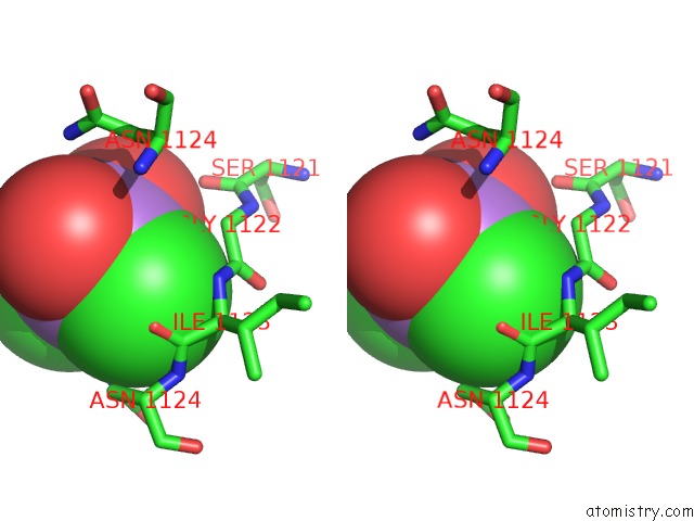
Arsenic binding site 4 out of 6 in 4u2v
Go back to
Arsenic binding site 4 out
of 6 in the Bak BH3-in-Groove Dimer (Gfp)
Mono view
Stereo pair view
Mono view
Stereo pair view
A full contact list of Arsenic with other atoms in the As binding
site number 4 of Bak BH3-in-Groove Dimer (Gfp) within 5.0Å range:
|
Arsenic binding site 5 out of 6 in 4u2v
Go back to
Arsenic binding site 5 out
of 6 in the Bak BH3-in-Groove Dimer (Gfp)
Mono view
Stereo pair view
Mono view
Stereo pair view
A full contact list of Arsenic with other atoms in the As binding
site number 5 of Bak BH3-in-Groove Dimer (Gfp) within 5.0Å range:
|
Arsenic binding site 6 out of 6 in 4u2v
Go back to
Arsenic binding site 6 out
of 6 in the Bak BH3-in-Groove Dimer (Gfp)
Mono view
Stereo pair view
Mono view
Stereo pair view
A full contact list of Arsenic with other atoms in the As binding
site number 6 of Bak BH3-in-Groove Dimer (Gfp) within 5.0Å range:
|
Reference:
J.M.Brouwer,
D.Westphal,
G.Dewson,
A.Y.Robin,
R.T.Uren,
R.Bartolo,
G.V.Thompson,
P.M.Colman,
R.M.Kluck,
P.E.Czabotar.
Bak Core and Latch Domains Separate During Activation, and Freed Core Domains Form Symmetric Homodimers. Mol.Cell V. 55 938 2014.
ISSN: ISSN 1097-2765
PubMed: 25175025
DOI: 10.1016/J.MOLCEL.2014.07.016
Page generated: Wed Jul 10 12:27:24 2024
ISSN: ISSN 1097-2765
PubMed: 25175025
DOI: 10.1016/J.MOLCEL.2014.07.016
Last articles
Zn in 9MJ5Zn in 9HNW
Zn in 9G0L
Zn in 9FNE
Zn in 9DZN
Zn in 9E0I
Zn in 9D32
Zn in 9DAK
Zn in 8ZXC
Zn in 8ZUF