Arsenic »
PDB 4zt2-5hr7 »
5cnx »
Arsenic in PDB 5cnx: Crystal Structure of Xaa-Pro Aminopeptidase From Escherichia Coli K12
Protein crystallography data
The structure of Crystal Structure of Xaa-Pro Aminopeptidase From Escherichia Coli K12, PDB code: 5cnx
was solved by
A.Kumar,
V.Are,
B.Ghosh,
S.Jamdar,
R.D.Makde,
with X-Ray Crystallography technique. A brief refinement statistics is given in the table below:
| Resolution Low / High (Å) | 48.90 / 2.60 |
| Space group | P 32 2 1 |
| Cell size a, b, c (Å), α, β, γ (°) | 224.202, 224.202, 74.636, 90.00, 90.00, 120.00 |
| R / Rfree (%) | 21.9 / 24 |
Other elements in 5cnx:
The structure of Crystal Structure of Xaa-Pro Aminopeptidase From Escherichia Coli K12 also contains other interesting chemical elements:
| Zinc | (Zn) | 6 atoms |
| Sodium | (Na) | 1 atom |
Arsenic Binding Sites:
The binding sites of Arsenic atom in the Crystal Structure of Xaa-Pro Aminopeptidase From Escherichia Coli K12
(pdb code 5cnx). This binding sites where shown within
5.0 Angstroms radius around Arsenic atom.
In total 3 binding sites of Arsenic where determined in the Crystal Structure of Xaa-Pro Aminopeptidase From Escherichia Coli K12, PDB code: 5cnx:
Jump to Arsenic binding site number: 1; 2; 3;
In total 3 binding sites of Arsenic where determined in the Crystal Structure of Xaa-Pro Aminopeptidase From Escherichia Coli K12, PDB code: 5cnx:
Jump to Arsenic binding site number: 1; 2; 3;
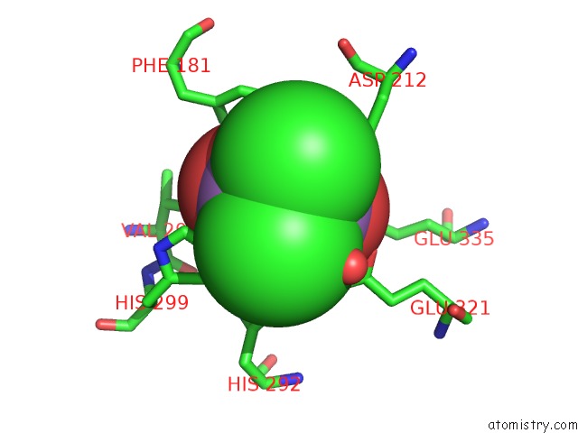
Arsenic binding site 1 out of 3 in 5cnx
Go back to
Arsenic binding site 1 out
of 3 in the Crystal Structure of Xaa-Pro Aminopeptidase From Escherichia Coli K12

Mono view

Stereo pair view
Mono view
Stereo pair view
A full contact list of Arsenic with other atoms in the As binding
site number 1 of Crystal Structure of Xaa-Pro Aminopeptidase From Escherichia Coli K12 within 5.0Å range:
|
Arsenic binding site 2 out of 3 in 5cnx
Go back to
Arsenic binding site 2 out
of 3 in the Crystal Structure of Xaa-Pro Aminopeptidase From Escherichia Coli K12
Mono view

Stereo pair view
Mono view
Stereo pair view
A full contact list of Arsenic with other atoms in the As binding
site number 2 of Crystal Structure of Xaa-Pro Aminopeptidase From Escherichia Coli K12 within 5.0Å range:
|
Arsenic binding site 3 out of 3 in 5cnx
Go back to
Arsenic binding site 3 out
of 3 in the Crystal Structure of Xaa-Pro Aminopeptidase From Escherichia Coli K12

Mono view

Stereo pair view
Mono view
Stereo pair view
A full contact list of Arsenic with other atoms in the As binding
site number 3 of Crystal Structure of Xaa-Pro Aminopeptidase From Escherichia Coli K12 within 5.0Å range:
|
Reference:
V.N.Are,
A.Kumar,
V.D.Goyal,
S.S.Gotad,
B.Ghosh,
R.Gadre,
S.N.Jamdar,
R.D.Makde.
Structures and Activities of Widely Conserved Small Prokaryotic Aminopeptidases-P Clarify Classification of M24B Peptidases Proteins 2018.
ISSN: ESSN 1097-0134
PubMed: 30536999
DOI: 10.1002/PROT.25641
Page generated: Wed Jul 10 12:46:12 2024
ISSN: ESSN 1097-0134
PubMed: 30536999
DOI: 10.1002/PROT.25641
Last articles
Zn in 9MJ5Zn in 9HNW
Zn in 9G0L
Zn in 9FNE
Zn in 9DZN
Zn in 9E0I
Zn in 9D32
Zn in 9DAK
Zn in 8ZXC
Zn in 8ZUF