Arsenic »
PDB 4zt2-5hr7 »
5e66 »
Arsenic in PDB 5e66: The Complex Structure of Hemagglutinin-Esterase-Fusion Mutant Protein From the Influenza D Virus with Receptor Analog 9-N-Ac-Sia
Protein crystallography data
The structure of The Complex Structure of Hemagglutinin-Esterase-Fusion Mutant Protein From the Influenza D Virus with Receptor Analog 9-N-Ac-Sia, PDB code: 5e66
was solved by
H.Song,
J.Qi,
Y.Shi,
G.F.Gao,
with X-Ray Crystallography technique. A brief refinement statistics is given in the table below:
| Resolution Low / High (Å) | 38.96 / 3.10 |
| Space group | I 21 3 |
| Cell size a, b, c (Å), α, β, γ (°) | 165.297, 165.297, 165.297, 90.00, 90.00, 90.00 |
| R / Rfree (%) | 23.4 / 26.5 |
Arsenic Binding Sites:
The binding sites of Arsenic atom in the The Complex Structure of Hemagglutinin-Esterase-Fusion Mutant Protein From the Influenza D Virus with Receptor Analog 9-N-Ac-Sia
(pdb code 5e66). This binding sites where shown within
5.0 Angstroms radius around Arsenic atom.
In total only one binding site of Arsenic was determined in the The Complex Structure of Hemagglutinin-Esterase-Fusion Mutant Protein From the Influenza D Virus with Receptor Analog 9-N-Ac-Sia, PDB code: 5e66:
In total only one binding site of Arsenic was determined in the The Complex Structure of Hemagglutinin-Esterase-Fusion Mutant Protein From the Influenza D Virus with Receptor Analog 9-N-Ac-Sia, PDB code: 5e66:
Arsenic binding site 1 out of 1 in 5e66
Go back to
Arsenic binding site 1 out
of 1 in the The Complex Structure of Hemagglutinin-Esterase-Fusion Mutant Protein From the Influenza D Virus with Receptor Analog 9-N-Ac-Sia
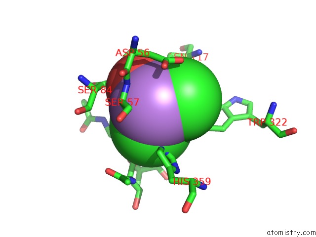
Mono view
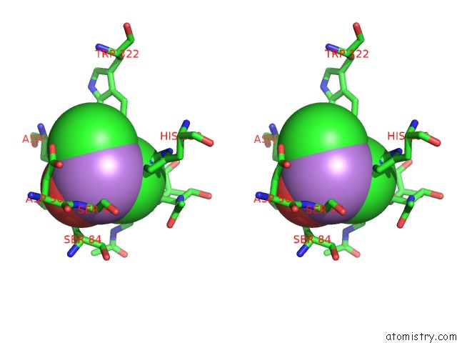
Stereo pair view
Mono view
Stereo pair view
A full contact list of Arsenic with other atoms in the As binding
site number 1 of The Complex Structure of Hemagglutinin-Esterase-Fusion Mutant Protein From the Influenza D Virus with Receptor Analog 9-N-Ac-Sia within 5.0Å range:
|
Reference:
H.Song,
J.Qi,
Z.Khedri,
S.Diaz,
H.Yu,
X.Chen,
A.Varki,
Y.Shi,
G.F.Gao.
An Open Receptor-Binding Cavity of Hemagglutinin-Esterase-Fusion Glycoprotein From Newly-Identified Influenza D Virus: Basis For Its Broad Cell Tropism Plos Pathog. V. 12 05411 2016.
ISSN: ESSN 1553-7374
PubMed: 26816272
DOI: 10.1371/JOURNAL.PPAT.1005411
Page generated: Wed Jul 10 12:47:47 2024
ISSN: ESSN 1553-7374
PubMed: 26816272
DOI: 10.1371/JOURNAL.PPAT.1005411
Last articles
Zn in 9MJ5Zn in 9HNW
Zn in 9G0L
Zn in 9FNE
Zn in 9DZN
Zn in 9E0I
Zn in 9D32
Zn in 9DAK
Zn in 8ZXC
Zn in 8ZUF