Arsenic »
PDB 5hrn-5o8x »
5hxd »
Arsenic in PDB 5hxd: Crystal Structure of Murein-Tripeptide Amidase Mpaa From Escherichia Coli O157
Protein crystallography data
The structure of Crystal Structure of Murein-Tripeptide Amidase Mpaa From Escherichia Coli O157, PDB code: 5hxd
was solved by
Y.Ma,
G.Bai,
X.Zhang,
J.Zhao,
Z.Yuan,
X.Kang,
Z.Li,
S.Mu,
X.Liu,
with X-Ray Crystallography technique. A brief refinement statistics is given in the table below:
| Resolution Low / High (Å) | 33.24 / 2.60 |
| Space group | P 31 |
| Cell size a, b, c (Å), α, β, γ (°) | 59.893, 59.893, 129.870, 90.00, 90.00, 120.00 |
| R / Rfree (%) | 17.2 / 24 |
Other elements in 5hxd:
The structure of Crystal Structure of Murein-Tripeptide Amidase Mpaa From Escherichia Coli O157 also contains other interesting chemical elements:
| Zinc | (Zn) | 2 atoms |
Arsenic Binding Sites:
The binding sites of Arsenic atom in the Crystal Structure of Murein-Tripeptide Amidase Mpaa From Escherichia Coli O157
(pdb code 5hxd). This binding sites where shown within
5.0 Angstroms radius around Arsenic atom.
In total 2 binding sites of Arsenic where determined in the Crystal Structure of Murein-Tripeptide Amidase Mpaa From Escherichia Coli O157, PDB code: 5hxd:
Jump to Arsenic binding site number: 1; 2;
In total 2 binding sites of Arsenic where determined in the Crystal Structure of Murein-Tripeptide Amidase Mpaa From Escherichia Coli O157, PDB code: 5hxd:
Jump to Arsenic binding site number: 1; 2;
Arsenic binding site 1 out of 2 in 5hxd
Go back to
Arsenic binding site 1 out
of 2 in the Crystal Structure of Murein-Tripeptide Amidase Mpaa From Escherichia Coli O157
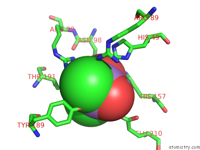
Mono view
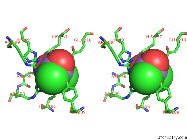
Stereo pair view
Mono view
Stereo pair view
A full contact list of Arsenic with other atoms in the As binding
site number 1 of Crystal Structure of Murein-Tripeptide Amidase Mpaa From Escherichia Coli O157 within 5.0Å range:
|
Arsenic binding site 2 out of 2 in 5hxd
Go back to
Arsenic binding site 2 out
of 2 in the Crystal Structure of Murein-Tripeptide Amidase Mpaa From Escherichia Coli O157
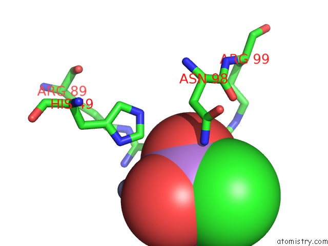
Mono view
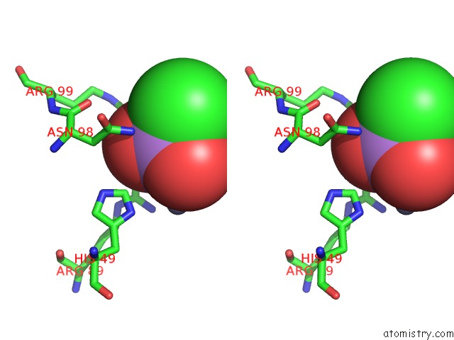
Stereo pair view
Mono view
Stereo pair view
A full contact list of Arsenic with other atoms in the As binding
site number 2 of Crystal Structure of Murein-Tripeptide Amidase Mpaa From Escherichia Coli O157 within 5.0Å range:
|
Reference:
Y.Ma,
G.Bai,
Y.Cui,
J.Zhao,
Z.Yuan,
X.Liu.
Crystal Structure of Murein-Tripeptide Amidase Mpaa From Escherichia Coli O157 at 2.6 Angstrom Resolution Protein Pept.Lett. V. 24 181 2017.
ISSN: ISSN 0929-8665
PubMed: 27894248
DOI: 10.2174/0929866523666161128153128
Page generated: Mon Jul 7 00:17:29 2025
ISSN: ISSN 0929-8665
PubMed: 27894248
DOI: 10.2174/0929866523666161128153128
Last articles
F in 7G5VF in 7G5W
F in 7G5U
F in 7G59
F in 7G5T
F in 7G5L
F in 7G5R
F in 7G5Q
F in 7G5P
F in 7G5M