Arsenic »
PDB 5oc4-5yva »
5xtu »
Arsenic in PDB 5xtu: Crystal Structure of Gdsl Esterase of Photobacterium Sp. J15
Protein crystallography data
The structure of Crystal Structure of Gdsl Esterase of Photobacterium Sp. J15, PDB code: 5xtu
was solved by
S.N.H.S.Mazlan,
M.A.Jonet,
T.C.Leow,
M.S.M.Ali,
R.N.Z.R.A.Rahman,
with X-Ray Crystallography technique. A brief refinement statistics is given in the table below:
| Resolution Low / High (Å) | 56.23 / 1.38 |
| Space group | P 21 21 21 |
| Cell size a, b, c (Å), α, β, γ (°) | 49.182, 66.461, 105.468, 90.00, 90.00, 90.00 |
| R / Rfree (%) | 15.8 / 17.9 |
Other elements in 5xtu:
The structure of Crystal Structure of Gdsl Esterase of Photobacterium Sp. J15 also contains other interesting chemical elements:
| Calcium | (Ca) | 1 atom |
| Chlorine | (Cl) | 1 atom |
Arsenic Binding Sites:
The binding sites of Arsenic atom in the Crystal Structure of Gdsl Esterase of Photobacterium Sp. J15
(pdb code 5xtu). This binding sites where shown within
5.0 Angstroms radius around Arsenic atom.
In total 2 binding sites of Arsenic where determined in the Crystal Structure of Gdsl Esterase of Photobacterium Sp. J15, PDB code: 5xtu:
Jump to Arsenic binding site number: 1; 2;
In total 2 binding sites of Arsenic where determined in the Crystal Structure of Gdsl Esterase of Photobacterium Sp. J15, PDB code: 5xtu:
Jump to Arsenic binding site number: 1; 2;
Arsenic binding site 1 out of 2 in 5xtu
Go back to
Arsenic binding site 1 out
of 2 in the Crystal Structure of Gdsl Esterase of Photobacterium Sp. J15
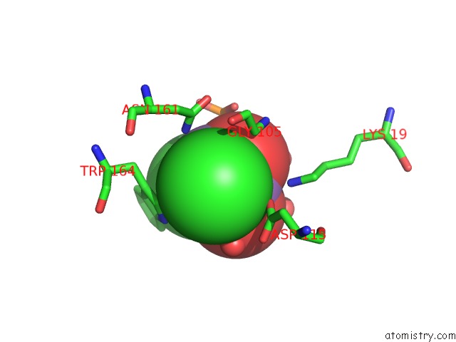
Mono view
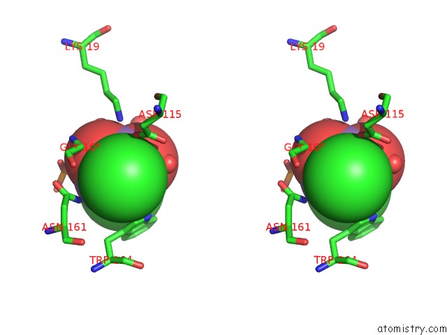
Stereo pair view
Mono view
Stereo pair view
A full contact list of Arsenic with other atoms in the As binding
site number 1 of Crystal Structure of Gdsl Esterase of Photobacterium Sp. J15 within 5.0Å range:
|
Arsenic binding site 2 out of 2 in 5xtu
Go back to
Arsenic binding site 2 out
of 2 in the Crystal Structure of Gdsl Esterase of Photobacterium Sp. J15
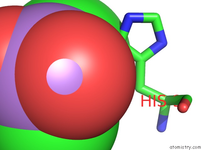
Mono view
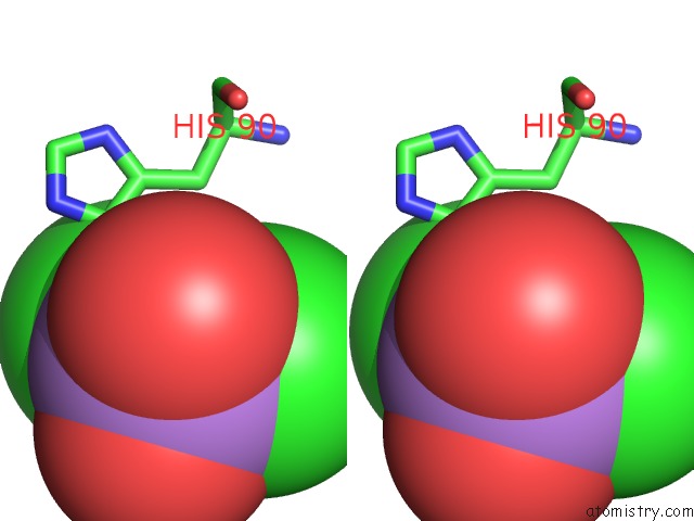
Stereo pair view
Mono view
Stereo pair view
A full contact list of Arsenic with other atoms in the As binding
site number 2 of Crystal Structure of Gdsl Esterase of Photobacterium Sp. J15 within 5.0Å range:
|
Reference:
S.N.H.S.Mazlan,
M.S.M.Ali,
R.N.Z.R.A.Rahman,
S.Sabri,
M.A.Jonet,
T.C.Leow.
Crystallization and Structure Elucidation of Gdsl Esterase of Photobacterium Sp. J15. Int. J. Biol. Macromol. V. 119 1188 2018.
ISSN: ISSN 1879-0003
PubMed: 30102982
DOI: 10.1016/J.IJBIOMAC.2018.08.022
Page generated: Wed Jul 10 13:11:57 2024
ISSN: ISSN 1879-0003
PubMed: 30102982
DOI: 10.1016/J.IJBIOMAC.2018.08.022
Last articles
Zn in 9MJ5Zn in 9HNW
Zn in 9G0L
Zn in 9FNE
Zn in 9DZN
Zn in 9E0I
Zn in 9D32
Zn in 9DAK
Zn in 8ZXC
Zn in 8ZUF