Arsenic »
PDB 6xna-7ji7 »
6zyu »
Arsenic in PDB 6zyu: Structure of the GLUA2 Ligand-Binding Domain (L483Y-N754S) in Complex with Glutamate and BPAM549
Protein crystallography data
The structure of Structure of the GLUA2 Ligand-Binding Domain (L483Y-N754S) in Complex with Glutamate and BPAM549, PDB code: 6zyu
was solved by
J.Dorosz,
K.M.Christensen,
J.S.Kastrup,
with X-Ray Crystallography technique. A brief refinement statistics is given in the table below:
| Resolution Low / High (Å) | 49.09 / 1.90 |
| Space group | P 21 21 2 |
| Cell size a, b, c (Å), α, β, γ (°) | 114.324, 163.084, 47.449, 90, 90, 90 |
| R / Rfree (%) | 16.2 / 18.7 |
Other elements in 6zyu:
The structure of Structure of the GLUA2 Ligand-Binding Domain (L483Y-N754S) in Complex with Glutamate and BPAM549 also contains other interesting chemical elements:
| Chlorine | (Cl) | 5 atoms |
| Fluorine | (F) | 6 atoms |
| Zinc | (Zn) | 8 atoms |
Arsenic Binding Sites:
The binding sites of Arsenic atom in the Structure of the GLUA2 Ligand-Binding Domain (L483Y-N754S) in Complex with Glutamate and BPAM549
(pdb code 6zyu). This binding sites where shown within
5.0 Angstroms radius around Arsenic atom.
In total only one binding site of Arsenic was determined in the Structure of the GLUA2 Ligand-Binding Domain (L483Y-N754S) in Complex with Glutamate and BPAM549, PDB code: 6zyu:
In total only one binding site of Arsenic was determined in the Structure of the GLUA2 Ligand-Binding Domain (L483Y-N754S) in Complex with Glutamate and BPAM549, PDB code: 6zyu:
Arsenic binding site 1 out of 1 in 6zyu
Go back to
Arsenic binding site 1 out
of 1 in the Structure of the GLUA2 Ligand-Binding Domain (L483Y-N754S) in Complex with Glutamate and BPAM549
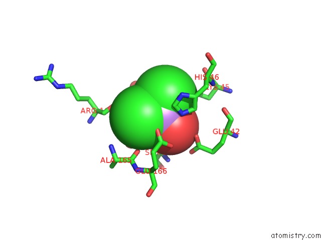
Mono view
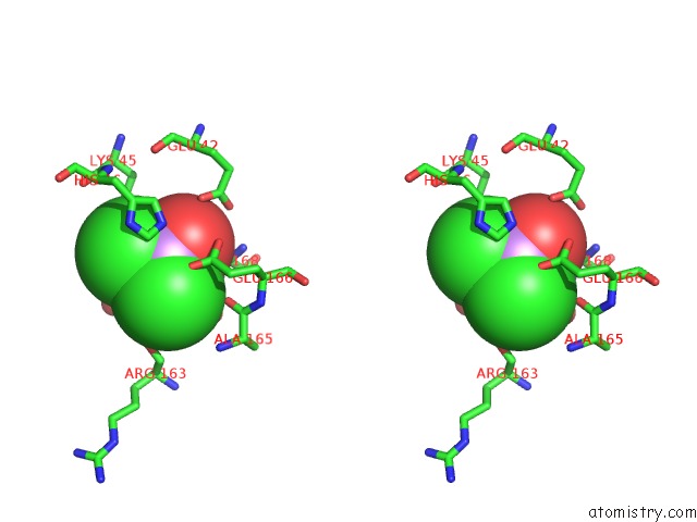
Stereo pair view
Mono view
Stereo pair view
A full contact list of Arsenic with other atoms in the As binding
site number 1 of Structure of the GLUA2 Ligand-Binding Domain (L483Y-N754S) in Complex with Glutamate and BPAM549 within 5.0Å range:
|
Reference:
J.Dorosz,
K.M.Christensen,
J.S.Kastrup.
Structure of the GLUA2 Ligand-Binding Domain (L483Y-N754S) in Complex with Glutamate and BPAM549 To Be Published.
Page generated: Mon Jul 7 00:53:30 2025
Last articles
Fe in 2YXOFe in 2YRS
Fe in 2YXC
Fe in 2YNM
Fe in 2YVJ
Fe in 2YP1
Fe in 2YU2
Fe in 2YU1
Fe in 2YQB
Fe in 2YOO