Arsenic »
PDB 6xna-7ji7 »
7dhy »
Arsenic in PDB 7dhy: Arsenic-Bound P53 Dna-Binding Domain Mutant G245S
Protein crystallography data
The structure of Arsenic-Bound P53 Dna-Binding Domain Mutant G245S, PDB code: 7dhy
was solved by
S.Chen,
M.Lu,
with X-Ray Crystallography technique. A brief refinement statistics is given in the table below:
| Resolution Low / High (Å) | 48.21 / 2.15 |
| Space group | P 1 21 1 |
| Cell size a, b, c (Å), α, β, γ (°) | 68.109, 68.243, 83.621, 90, 90.03, 90 |
| R / Rfree (%) | 18.7 / 21.9 |
Other elements in 7dhy:
The structure of Arsenic-Bound P53 Dna-Binding Domain Mutant G245S also contains other interesting chemical elements:
| Zinc | (Zn) | 4 atoms |
Arsenic Binding Sites:
The binding sites of Arsenic atom in the Arsenic-Bound P53 Dna-Binding Domain Mutant G245S
(pdb code 7dhy). This binding sites where shown within
5.0 Angstroms radius around Arsenic atom.
In total 4 binding sites of Arsenic where determined in the Arsenic-Bound P53 Dna-Binding Domain Mutant G245S, PDB code: 7dhy:
Jump to Arsenic binding site number: 1; 2; 3; 4;
In total 4 binding sites of Arsenic where determined in the Arsenic-Bound P53 Dna-Binding Domain Mutant G245S, PDB code: 7dhy:
Jump to Arsenic binding site number: 1; 2; 3; 4;
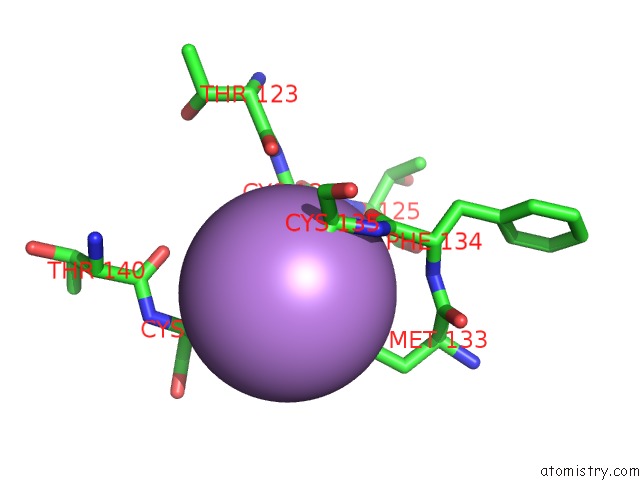
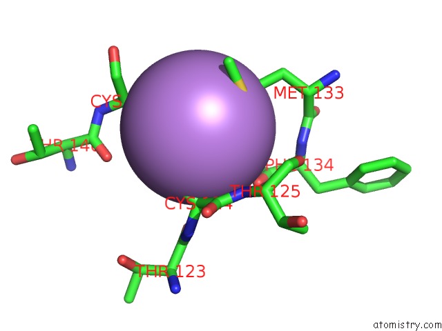
Arsenic binding site 1 out of 4 in 7dhy
Go back to
Arsenic binding site 1 out
of 4 in the Arsenic-Bound P53 Dna-Binding Domain Mutant G245S
Mono view
Stereo pair view
Mono view
Stereo pair view
A full contact list of Arsenic with other atoms in the As binding
site number 1 of Arsenic-Bound P53 Dna-Binding Domain Mutant G245S within 5.0Å range:
|
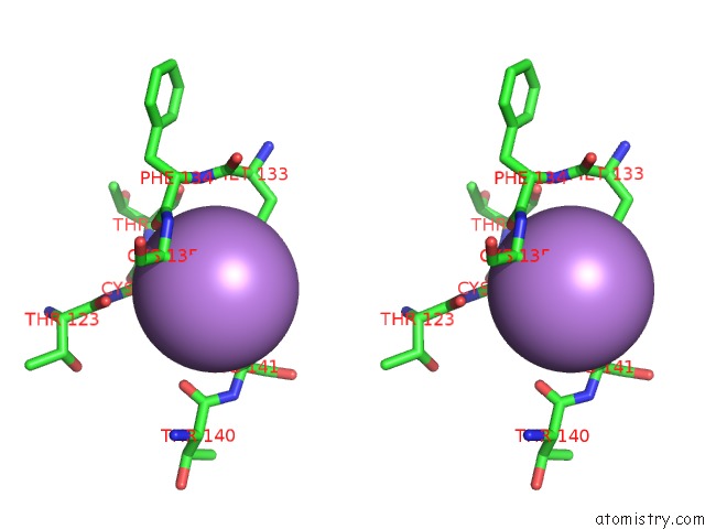
Arsenic binding site 2 out of 4 in 7dhy
Go back to
Arsenic binding site 2 out
of 4 in the Arsenic-Bound P53 Dna-Binding Domain Mutant G245S

Mono view
Stereo pair view
Mono view
Stereo pair view
A full contact list of Arsenic with other atoms in the As binding
site number 2 of Arsenic-Bound P53 Dna-Binding Domain Mutant G245S within 5.0Å range:
|
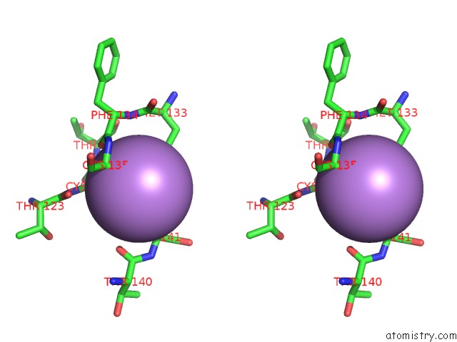
Arsenic binding site 3 out of 4 in 7dhy
Go back to
Arsenic binding site 3 out
of 4 in the Arsenic-Bound P53 Dna-Binding Domain Mutant G245S

Mono view
Stereo pair view
Mono view
Stereo pair view
A full contact list of Arsenic with other atoms in the As binding
site number 3 of Arsenic-Bound P53 Dna-Binding Domain Mutant G245S within 5.0Å range:
|
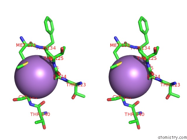
Arsenic binding site 4 out of 4 in 7dhy
Go back to
Arsenic binding site 4 out
of 4 in the Arsenic-Bound P53 Dna-Binding Domain Mutant G245S
Mono view

Stereo pair view
Mono view
Stereo pair view
A full contact list of Arsenic with other atoms in the As binding
site number 4 of Arsenic-Bound P53 Dna-Binding Domain Mutant G245S within 5.0Å range:
|
Reference:
S.Chen,
J.L.Wu,
Y.Liang,
Y.G.Tang,
H.X.Song,
L.L.Wu,
Y.F.Xing,
N.Yan,
Y.T.Li,
Z.Y.Wang,
S.J.Xiao,
X.Lu,
S.J.Chen,
M.Lu.
Arsenic Trioxide Rescues Structural P53 Mutations Through A Cryptic Allosteric Site. Cancer Cell V. 39 225 2021.
ISSN: ISSN 1535-6108
PubMed: 33357454
DOI: 10.1016/J.CCELL.2020.11.013
Page generated: Mon Jul 7 00:55:39 2025
ISSN: ISSN 1535-6108
PubMed: 33357454
DOI: 10.1016/J.CCELL.2020.11.013
Last articles
Br in 2BYDBr in 2CEJ
Br in 2C69
Br in 2B4G
Br in 2C68
Br in 2A9W
Br in 2BU1
Br in 2BHE
Br in 2B63
Br in 2AZ2