Arsenic »
PDB 1z6b-2im2 »
2dyb »
Arsenic in PDB 2dyb: The Crystal Structure of Human P40(Phox)
Protein crystallography data
The structure of The Crystal Structure of Human P40(Phox), PDB code: 2dyb
was solved by
K.Honbou,
with X-Ray Crystallography technique. A brief refinement statistics is given in the table below:
| Resolution Low / High (Å) | 40.80 / 3.15 |
| Space group | C 2 2 21 |
| Cell size a, b, c (Å), α, β, γ (°) | 146.273, 189.813, 79.883, 90.00, 90.00, 90.00 |
| R / Rfree (%) | 26.3 / 30.2 |
Arsenic Binding Sites:
The binding sites of Arsenic atom in the The Crystal Structure of Human P40(Phox)
(pdb code 2dyb). This binding sites where shown within
5.0 Angstroms radius around Arsenic atom.
In total 2 binding sites of Arsenic where determined in the The Crystal Structure of Human P40(Phox), PDB code: 2dyb:
Jump to Arsenic binding site number: 1; 2;
In total 2 binding sites of Arsenic where determined in the The Crystal Structure of Human P40(Phox), PDB code: 2dyb:
Jump to Arsenic binding site number: 1; 2;
Arsenic binding site 1 out of 2 in 2dyb
Go back to
Arsenic binding site 1 out
of 2 in the The Crystal Structure of Human P40(Phox)
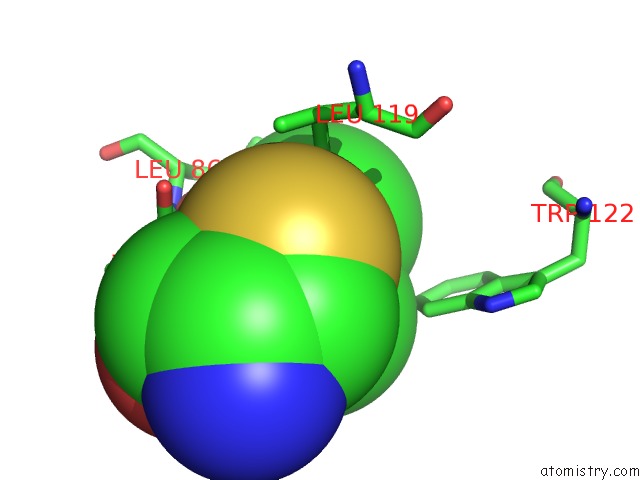
Mono view
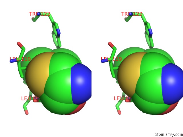
Stereo pair view
Mono view
Stereo pair view
A full contact list of Arsenic with other atoms in the As binding
site number 1 of The Crystal Structure of Human P40(Phox) within 5.0Å range:
|
Arsenic binding site 2 out of 2 in 2dyb
Go back to
Arsenic binding site 2 out
of 2 in the The Crystal Structure of Human P40(Phox)
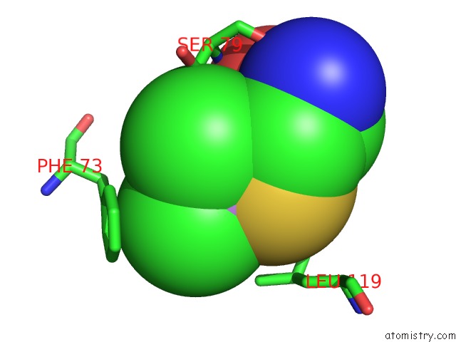
Mono view
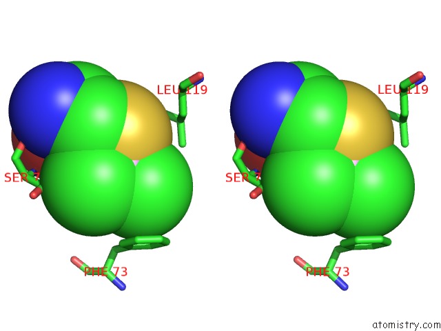
Stereo pair view
Mono view
Stereo pair view
A full contact list of Arsenic with other atoms in the As binding
site number 2 of The Crystal Structure of Human P40(Phox) within 5.0Å range:
|
Reference:
K.Honbou,
R.Minakami,
S.Yuzawa,
R.Takeya,
N.N.Suzuki,
S.Kamakura,
H.Sumimoto,
F.Inagaki.
Full-Length P40PHOX Structure Suggests A Basis For Regulation Mechanism of Its Membrane Binding. Embo J. V. 26 1176 2007.
ISSN: ISSN 0261-4189
PubMed: 17290225
DOI: 10.1038/SJ.EMBOJ.7601561
Page generated: Sun Jul 6 23:07:25 2025
ISSN: ISSN 0261-4189
PubMed: 17290225
DOI: 10.1038/SJ.EMBOJ.7601561
Last articles
Mg in 4OOGMg in 4OOP
Mg in 4OMF
Mg in 4OO1
Mg in 4OKM
Mg in 4OLS
Mg in 4OL0
Mg in 4OKK
Mg in 4OKJ
Mg in 4OKQ