Arsenic »
PDB 1z6b-2im2 »
2e11 »
Arsenic in PDB 2e11: The Crystal Structure of XC1258 From Xanthomonas Campestris: A Cn- Hydrolase Superfamily Protein with An Arsenic Adduct in the Active Site
Protein crystallography data
The structure of The Crystal Structure of XC1258 From Xanthomonas Campestris: A Cn- Hydrolase Superfamily Protein with An Arsenic Adduct in the Active Site, PDB code: 2e11
was solved by
K.-H.Chin,
Y.-D.Tsai,
N.-L.Chan,
K.-F.Huang,
A.H.-J.Wang,
S.-H.Chou,
with X-Ray Crystallography technique. A brief refinement statistics is given in the table below:
| Resolution Low / High (Å) | 104.83 / 1.73 |
| Space group | P 21 21 2 |
| Cell size a, b, c (Å), α, β, γ (°) | 143.280, 154.305, 51.158, 90.00, 90.00, 90.00 |
| R / Rfree (%) | 14 / 21.7 |
Arsenic Binding Sites:
The binding sites of Arsenic atom in the The Crystal Structure of XC1258 From Xanthomonas Campestris: A Cn- Hydrolase Superfamily Protein with An Arsenic Adduct in the Active Site
(pdb code 2e11). This binding sites where shown within
5.0 Angstroms radius around Arsenic atom.
In total 4 binding sites of Arsenic where determined in the The Crystal Structure of XC1258 From Xanthomonas Campestris: A Cn- Hydrolase Superfamily Protein with An Arsenic Adduct in the Active Site, PDB code: 2e11:
Jump to Arsenic binding site number: 1; 2; 3; 4;
In total 4 binding sites of Arsenic where determined in the The Crystal Structure of XC1258 From Xanthomonas Campestris: A Cn- Hydrolase Superfamily Protein with An Arsenic Adduct in the Active Site, PDB code: 2e11:
Jump to Arsenic binding site number: 1; 2; 3; 4;
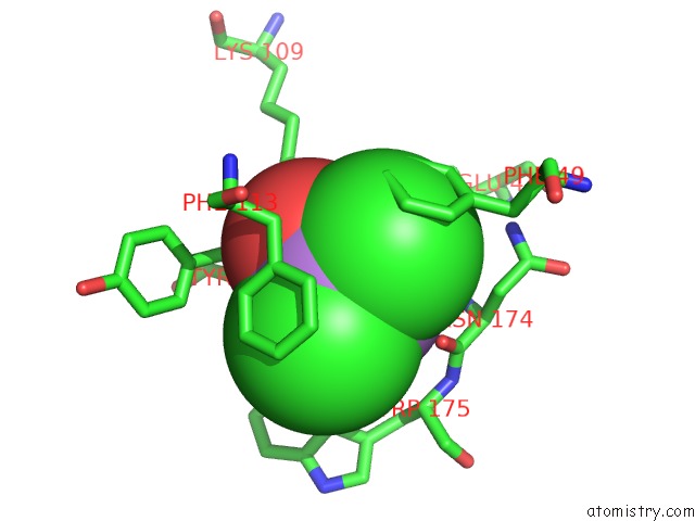
Arsenic binding site 1 out of 4 in 2e11
Go back to
Arsenic binding site 1 out
of 4 in the The Crystal Structure of XC1258 From Xanthomonas Campestris: A Cn- Hydrolase Superfamily Protein with An Arsenic Adduct in the Active Site

Mono view
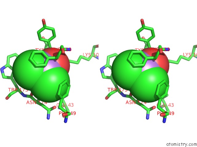
Stereo pair view
Mono view
Stereo pair view
A full contact list of Arsenic with other atoms in the As binding
site number 1 of The Crystal Structure of XC1258 From Xanthomonas Campestris: A Cn- Hydrolase Superfamily Protein with An Arsenic Adduct in the Active Site within 5.0Å range:
|
Arsenic binding site 2 out of 4 in 2e11
Go back to
Arsenic binding site 2 out
of 4 in the The Crystal Structure of XC1258 From Xanthomonas Campestris: A Cn- Hydrolase Superfamily Protein with An Arsenic Adduct in the Active Site
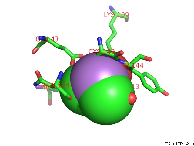
Mono view
Stereo pair view
Mono view
Stereo pair view
A full contact list of Arsenic with other atoms in the As binding
site number 2 of The Crystal Structure of XC1258 From Xanthomonas Campestris: A Cn- Hydrolase Superfamily Protein with An Arsenic Adduct in the Active Site within 5.0Å range:
|
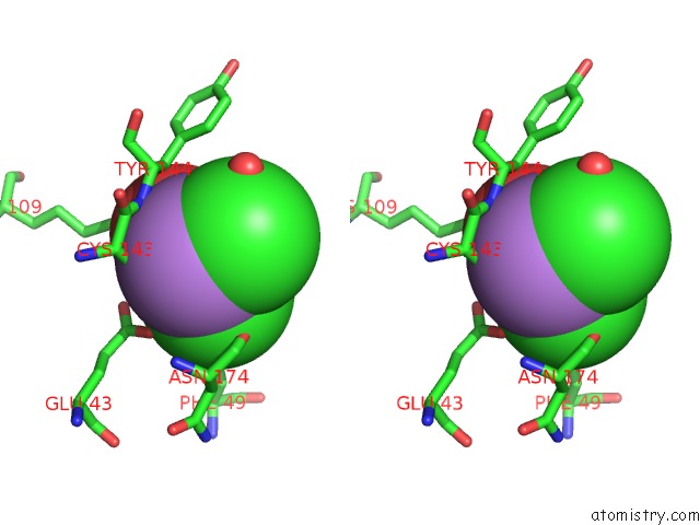
Arsenic binding site 3 out of 4 in 2e11
Go back to
Arsenic binding site 3 out
of 4 in the The Crystal Structure of XC1258 From Xanthomonas Campestris: A Cn- Hydrolase Superfamily Protein with An Arsenic Adduct in the Active Site
Mono view

Stereo pair view
Mono view
Stereo pair view
A full contact list of Arsenic with other atoms in the As binding
site number 3 of The Crystal Structure of XC1258 From Xanthomonas Campestris: A Cn- Hydrolase Superfamily Protein with An Arsenic Adduct in the Active Site within 5.0Å range:
|
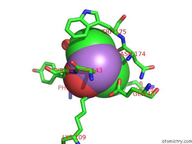
Arsenic binding site 4 out of 4 in 2e11
Go back to
Arsenic binding site 4 out
of 4 in the The Crystal Structure of XC1258 From Xanthomonas Campestris: A Cn- Hydrolase Superfamily Protein with An Arsenic Adduct in the Active Site
Mono view

Stereo pair view
Mono view
Stereo pair view
A full contact list of Arsenic with other atoms in the As binding
site number 4 of The Crystal Structure of XC1258 From Xanthomonas Campestris: A Cn- Hydrolase Superfamily Protein with An Arsenic Adduct in the Active Site within 5.0Å range:
|
Reference:
K.-H.Chin,
Y.-D.Tsai,
N.-L.Chan,
K.-F.Huang,
A.H.-J.Wang,
S.-H.Chou.
The Crystal Structure of XC1258 From Xanthomonas Campestris: A Putative Procaryotic Nit Protein with An Arsenic Adduct in the Active Site Proteins V. 69 665 2007.
ISSN: ISSN 0887-3585
PubMed: 17640068
DOI: 10.1002/PROT.21501
Page generated: Sun Jul 6 23:07:31 2025
ISSN: ISSN 0887-3585
PubMed: 17640068
DOI: 10.1002/PROT.21501
Last articles
Mg in 3X0EMg in 3X1L
Mg in 3X1D
Mg in 3WZY
Mg in 3WZX
Mg in 3WYM
Mg in 3WZW
Mg in 3WZV
Mg in 3WYL
Mg in 3WVL