Arsenic »
PDB 1z6b-2im2 »
2gbm »
Arsenic in PDB 2gbm: Crystal Structure of the 35-36 8 Glycine Insertion Mutant of Ubiquitin
Protein crystallography data
The structure of Crystal Structure of the 35-36 8 Glycine Insertion Mutant of Ubiquitin, PDB code: 2gbm
was solved by
D.M.Ferraro,
D.J.Ferraro,
S.Ramaswamy,
A.D.Robertson,
with X-Ray Crystallography technique. A brief refinement statistics is given in the table below:
| Resolution Low / High (Å) | 19.51 / 1.55 |
| Space group | P 21 21 21 |
| Cell size a, b, c (Å), α, β, γ (°) | 48.817, 49.786, 123.147, 90.00, 90.00, 90.00 |
| R / Rfree (%) | 21.3 / 22.7 |
Arsenic Binding Sites:
The binding sites of Arsenic atom in the Crystal Structure of the 35-36 8 Glycine Insertion Mutant of Ubiquitin
(pdb code 2gbm). This binding sites where shown within
5.0 Angstroms radius around Arsenic atom.
In total 3 binding sites of Arsenic where determined in the Crystal Structure of the 35-36 8 Glycine Insertion Mutant of Ubiquitin, PDB code: 2gbm:
Jump to Arsenic binding site number: 1; 2; 3;
In total 3 binding sites of Arsenic where determined in the Crystal Structure of the 35-36 8 Glycine Insertion Mutant of Ubiquitin, PDB code: 2gbm:
Jump to Arsenic binding site number: 1; 2; 3;
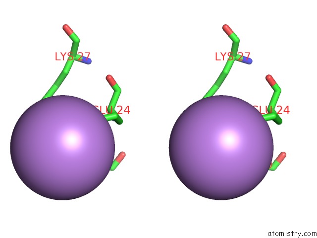
Arsenic binding site 1 out of 3 in 2gbm
Go back to
Arsenic binding site 1 out
of 3 in the Crystal Structure of the 35-36 8 Glycine Insertion Mutant of Ubiquitin
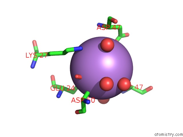
Mono view
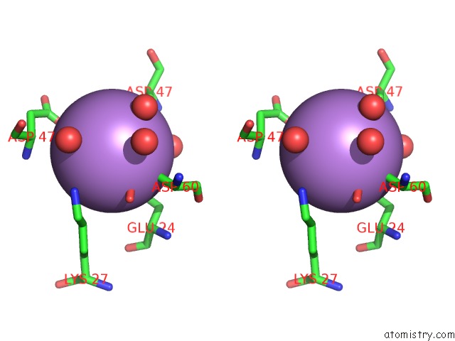
Stereo pair view
Mono view
Stereo pair view
A full contact list of Arsenic with other atoms in the As binding
site number 1 of Crystal Structure of the 35-36 8 Glycine Insertion Mutant of Ubiquitin within 5.0Å range:
|
Arsenic binding site 2 out of 3 in 2gbm
Go back to
Arsenic binding site 2 out
of 3 in the Crystal Structure of the 35-36 8 Glycine Insertion Mutant of Ubiquitin
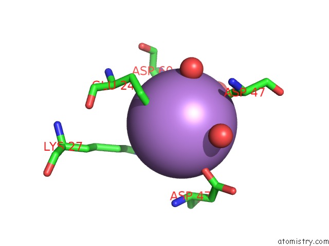
Mono view

Stereo pair view
Mono view
Stereo pair view
A full contact list of Arsenic with other atoms in the As binding
site number 2 of Crystal Structure of the 35-36 8 Glycine Insertion Mutant of Ubiquitin within 5.0Å range:
|
Arsenic binding site 3 out of 3 in 2gbm
Go back to
Arsenic binding site 3 out
of 3 in the Crystal Structure of the 35-36 8 Glycine Insertion Mutant of Ubiquitin
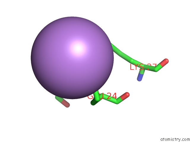
Mono view
Stereo pair view
Mono view
Stereo pair view
A full contact list of Arsenic with other atoms in the As binding
site number 3 of Crystal Structure of the 35-36 8 Glycine Insertion Mutant of Ubiquitin within 5.0Å range:
|
Reference:
D.M.Ferraro,
D.J.Ferraro,
S.Ramaswamy,
A.D.Robertson.
Structures of Ubiquitin Insertion Mutants Support Site-Specific Reflex Response to Insertions Hypothesis. J.Mol.Biol. V. 359 390 2006.
ISSN: ISSN 0022-2836
PubMed: 16647719
DOI: 10.1016/J.JMB.2006.03.047
Page generated: Sun Jul 6 23:08:34 2025
ISSN: ISSN 0022-2836
PubMed: 16647719
DOI: 10.1016/J.JMB.2006.03.047
Last articles
Mg in 6O3PMg in 6O2R
Mg in 6O2Q
Mg in 6O36
Mg in 6O2P
Mg in 6O1V
Mg in 6O0J
Mg in 6O1E
Mg in 6O0G
Mg in 6NYY