Arsenic »
PDB 2xod-3g2f »
3et6 »
Arsenic in PDB 3et6: The Crystal Structure of the Catalytic Domain of A Eukaryotic Guanylate Cyclase
Protein crystallography data
The structure of The Crystal Structure of the Catalytic Domain of A Eukaryotic Guanylate Cyclase, PDB code: 3et6
was solved by
J.A.Winger,
E.R.Derbyshire,
M.H.Lamers,
M.A.Marletta,
J.Kuriyan,
with X-Ray Crystallography technique. A brief refinement statistics is given in the table below:
| Resolution Low / High (Å) | 28.00 / 2.55 |
| Space group | P 32 2 1 |
| Cell size a, b, c (Å), α, β, γ (°) | 123.678, 123.678, 62.822, 90.00, 90.00, 120.00 |
| R / Rfree (%) | 17.2 / 21.5 |
Arsenic Binding Sites:
The binding sites of Arsenic atom in the The Crystal Structure of the Catalytic Domain of A Eukaryotic Guanylate Cyclase
(pdb code 3et6). This binding sites where shown within
5.0 Angstroms radius around Arsenic atom.
In total 5 binding sites of Arsenic where determined in the The Crystal Structure of the Catalytic Domain of A Eukaryotic Guanylate Cyclase, PDB code: 3et6:
Jump to Arsenic binding site number: 1; 2; 3; 4; 5;
In total 5 binding sites of Arsenic where determined in the The Crystal Structure of the Catalytic Domain of A Eukaryotic Guanylate Cyclase, PDB code: 3et6:
Jump to Arsenic binding site number: 1; 2; 3; 4; 5;
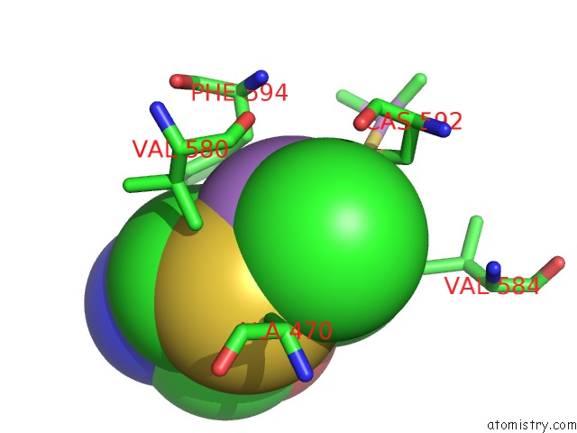
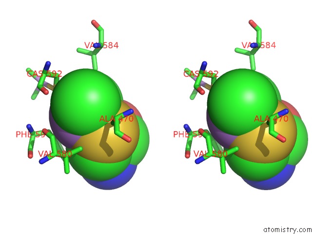
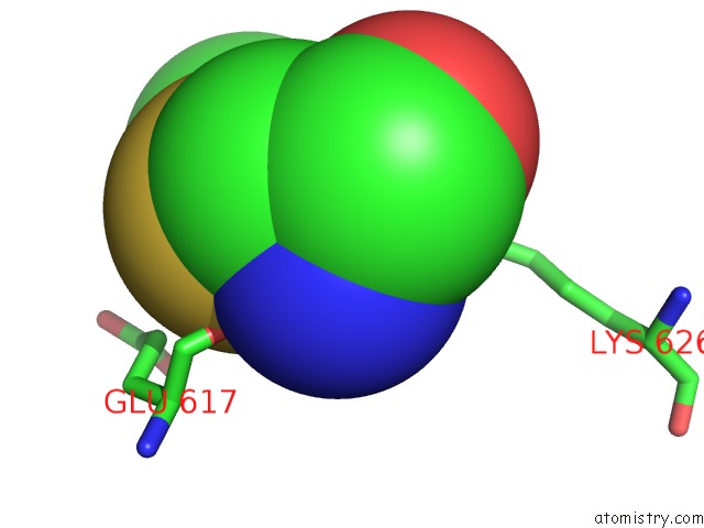
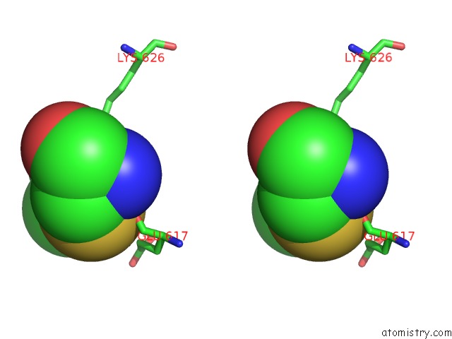
Arsenic binding site 1 out of 5 in 3et6
Go back to
Arsenic binding site 1 out
of 5 in the The Crystal Structure of the Catalytic Domain of A Eukaryotic Guanylate Cyclase
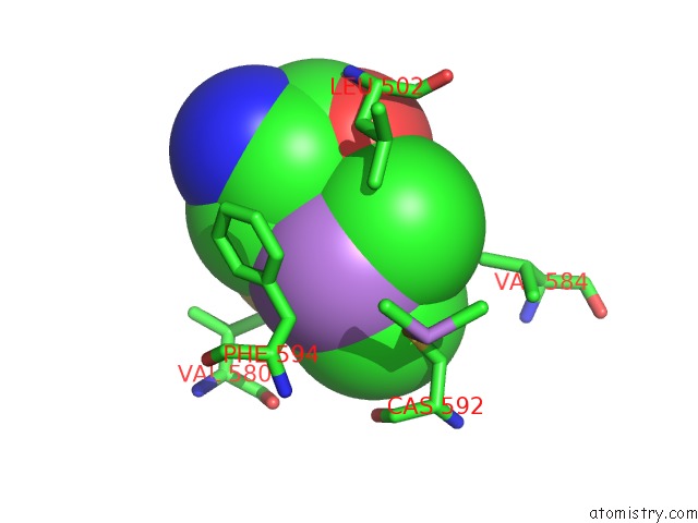
Mono view
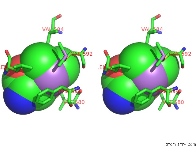
Stereo pair view
Mono view
Stereo pair view
A full contact list of Arsenic with other atoms in the As binding
site number 1 of The Crystal Structure of the Catalytic Domain of A Eukaryotic Guanylate Cyclase within 5.0Å range:
|
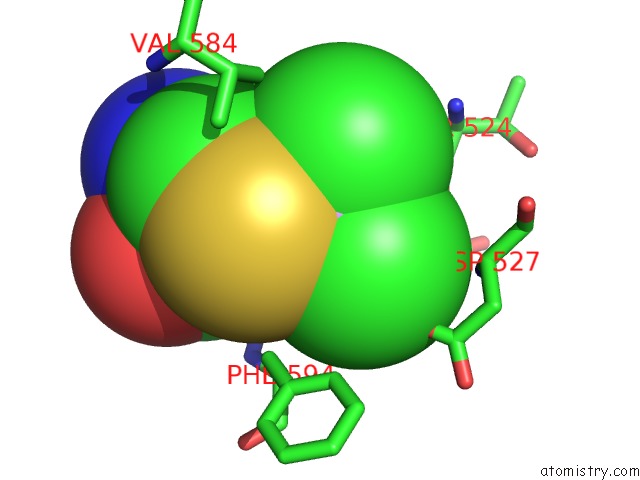
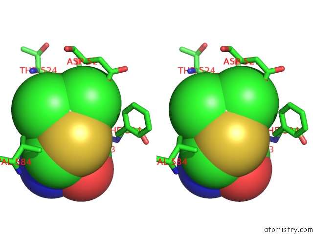
Arsenic binding site 2 out of 5 in 3et6
Go back to
Arsenic binding site 2 out
of 5 in the The Crystal Structure of the Catalytic Domain of A Eukaryotic Guanylate Cyclase
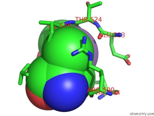
Mono view
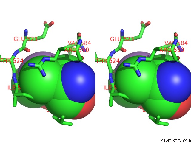
Stereo pair view
Mono view
Stereo pair view
A full contact list of Arsenic with other atoms in the As binding
site number 2 of The Crystal Structure of the Catalytic Domain of A Eukaryotic Guanylate Cyclase within 5.0Å range:
|
Arsenic binding site 3 out of 5 in 3et6
Go back to
Arsenic binding site 3 out
of 5 in the The Crystal Structure of the Catalytic Domain of A Eukaryotic Guanylate Cyclase
Mono view
Stereo pair view
Mono view
Stereo pair view
A full contact list of Arsenic with other atoms in the As binding
site number 3 of The Crystal Structure of the Catalytic Domain of A Eukaryotic Guanylate Cyclase within 5.0Å range:
|
Arsenic binding site 4 out of 5 in 3et6
Go back to
Arsenic binding site 4 out
of 5 in the The Crystal Structure of the Catalytic Domain of A Eukaryotic Guanylate Cyclase
Mono view
Stereo pair view
Mono view
Stereo pair view
A full contact list of Arsenic with other atoms in the As binding
site number 4 of The Crystal Structure of the Catalytic Domain of A Eukaryotic Guanylate Cyclase within 5.0Å range:
|
Arsenic binding site 5 out of 5 in 3et6
Go back to
Arsenic binding site 5 out
of 5 in the The Crystal Structure of the Catalytic Domain of A Eukaryotic Guanylate Cyclase
Mono view
Stereo pair view
Mono view
Stereo pair view
A full contact list of Arsenic with other atoms in the As binding
site number 5 of The Crystal Structure of the Catalytic Domain of A Eukaryotic Guanylate Cyclase within 5.0Å range:
|
Reference:
J.A.Winger,
E.R.Derbyshire,
M.H.Lamers,
M.A.Marletta,
J.Kuriyan.
The Crystal Structure of the Catalytic Domain of A Eukaryotic Guanylate Cyclase. Bmc Struct.Biol. V. 8 42 2008.
ISSN: ESSN 1472-6807
PubMed: 18842118
DOI: 10.1186/1472-6807-8-42
Page generated: Sun Jul 6 23:21:26 2025
ISSN: ESSN 1472-6807
PubMed: 18842118
DOI: 10.1186/1472-6807-8-42
Last articles
La in 6IP9La in 1DJG
La in 6DAM
La in 5KKB
La in 2OQR
La in 2RPV
La in 5KIJ
La in 2K0J
La in 2I18
La in 1KQV