Arsenic »
PDB 3g3s-3n5t »
3kzp »
Arsenic in PDB 3kzp: Crystal Structure of Putative Diguanylate Cyclase/Phosphodiesterase From Listaria Monocytigenes
Protein crystallography data
The structure of Crystal Structure of Putative Diguanylate Cyclase/Phosphodiesterase From Listaria Monocytigenes, PDB code: 3kzp
was solved by
M.M.Klimecka,
M.Chruszcz,
M.D.Zimmerman,
M.Kudritska,
A.Savchenko,
A.Edwards,
A.Joachimiak,
W.Minor,
Midwest Center For Structuralgenomics (Mcsg),
with X-Ray Crystallography technique. A brief refinement statistics is given in the table below:
| Resolution Low / High (Å) | 50.00 / 2.00 |
| Space group | P 21 21 21 |
| Cell size a, b, c (Å), α, β, γ (°) | 59.766, 92.040, 96.210, 90.00, 90.00, 90.00 |
| R / Rfree (%) | 18.5 / 23.9 |
Other elements in 3kzp:
The structure of Crystal Structure of Putative Diguanylate Cyclase/Phosphodiesterase From Listaria Monocytigenes also contains other interesting chemical elements:
| Calcium | (Ca) | 2 atoms |
| Chlorine | (Cl) | 6 atoms |
Arsenic Binding Sites:
The binding sites of Arsenic atom in the Crystal Structure of Putative Diguanylate Cyclase/Phosphodiesterase From Listaria Monocytigenes
(pdb code 3kzp). This binding sites where shown within
5.0 Angstroms radius around Arsenic atom.
In total 2 binding sites of Arsenic where determined in the Crystal Structure of Putative Diguanylate Cyclase/Phosphodiesterase From Listaria Monocytigenes, PDB code: 3kzp:
Jump to Arsenic binding site number: 1; 2;
In total 2 binding sites of Arsenic where determined in the Crystal Structure of Putative Diguanylate Cyclase/Phosphodiesterase From Listaria Monocytigenes, PDB code: 3kzp:
Jump to Arsenic binding site number: 1; 2;
Arsenic binding site 1 out of 2 in 3kzp
Go back to
Arsenic binding site 1 out
of 2 in the Crystal Structure of Putative Diguanylate Cyclase/Phosphodiesterase From Listaria Monocytigenes
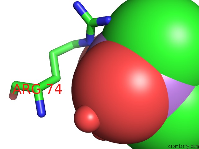
Mono view
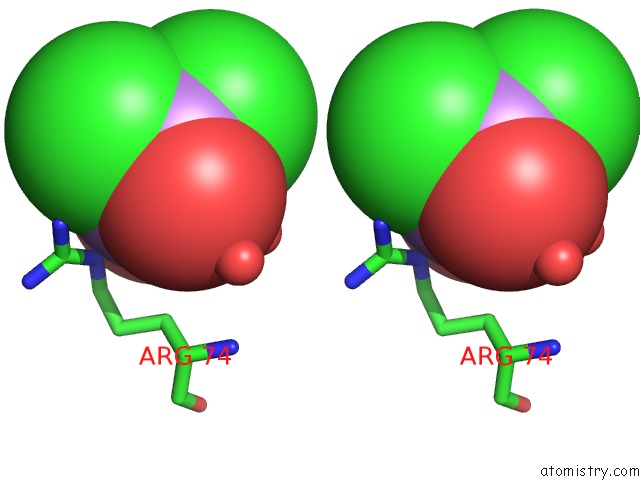
Stereo pair view
Mono view
Stereo pair view
A full contact list of Arsenic with other atoms in the As binding
site number 1 of Crystal Structure of Putative Diguanylate Cyclase/Phosphodiesterase From Listaria Monocytigenes within 5.0Å range:
|
Arsenic binding site 2 out of 2 in 3kzp
Go back to
Arsenic binding site 2 out
of 2 in the Crystal Structure of Putative Diguanylate Cyclase/Phosphodiesterase From Listaria Monocytigenes
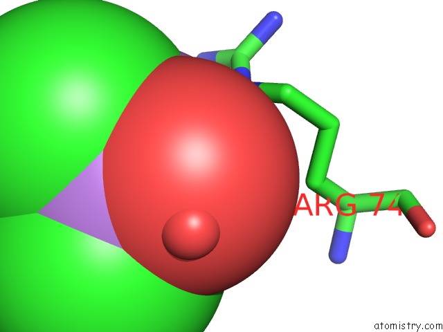
Mono view
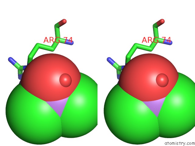
Stereo pair view
Mono view
Stereo pair view
A full contact list of Arsenic with other atoms in the As binding
site number 2 of Crystal Structure of Putative Diguanylate Cyclase/Phosphodiesterase From Listaria Monocytigenes within 5.0Å range:
|
Reference:
M.M.Klimecka,
M.Chruszcz,
M.D.Zimmerman,
M.Kudritska,
A.Savchenko,
A.Edwards,
A.Joachimiak,
W.Minor.
Crystal Structure of Putative Diguanylate Cyclase/Phosphodiesterase From Listaria Monocytigenes To Be Published.
Page generated: Sun Jul 6 23:26:55 2025
Last articles
Mg in 1HI8Mg in 1HJ6
Mg in 1HBN
Mg in 1HBU
Mg in 1HH4
Mg in 1HE8
Mg in 1HE1
Mg in 1HDI
Mg in 1HCK
Mg in 1HBO