Arsenic »
PDB 3g3s-3n5t »
3msz »
Arsenic in PDB 3msz: Crystal Structure of Glutaredoxin 1 From Francisella Tularensis Complexed with Cacodylate
Protein crystallography data
The structure of Crystal Structure of Glutaredoxin 1 From Francisella Tularensis Complexed with Cacodylate, PDB code: 3msz
was solved by
N.Maltseva,
Y.Kim,
K.Kwon,
W.F.Anderson,
A.Joachimiak,
with X-Ray Crystallography technique. A brief refinement statistics is given in the table below:
| Resolution Low / High (Å) | 40.72 / 2.05 |
| Space group | P 32 2 1 |
| Cell size a, b, c (Å), α, β, γ (°) | 49.139, 49.139, 140.199, 90.00, 90.00, 120.00 |
| R / Rfree (%) | 17.3 / 22.3 |
Arsenic Binding Sites:
The binding sites of Arsenic atom in the Crystal Structure of Glutaredoxin 1 From Francisella Tularensis Complexed with Cacodylate
(pdb code 3msz). This binding sites where shown within
5.0 Angstroms radius around Arsenic atom.
In total only one binding site of Arsenic was determined in the Crystal Structure of Glutaredoxin 1 From Francisella Tularensis Complexed with Cacodylate, PDB code: 3msz:
In total only one binding site of Arsenic was determined in the Crystal Structure of Glutaredoxin 1 From Francisella Tularensis Complexed with Cacodylate, PDB code: 3msz:
Arsenic binding site 1 out of 1 in 3msz
Go back to
Arsenic binding site 1 out
of 1 in the Crystal Structure of Glutaredoxin 1 From Francisella Tularensis Complexed with Cacodylate
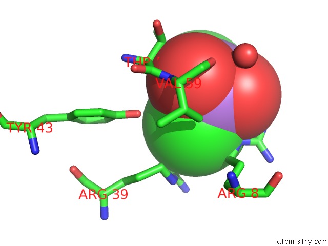
Mono view
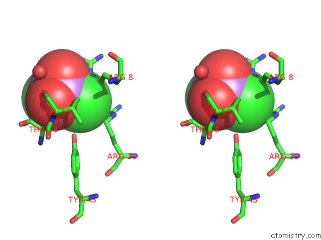
Stereo pair view
Mono view
Stereo pair view
A full contact list of Arsenic with other atoms in the As binding
site number 1 of Crystal Structure of Glutaredoxin 1 From Francisella Tularensis Complexed with Cacodylate within 5.0Å range:
|
Reference:
N.Maltseva,
Y.Kim,
K.Kwon,
W.F.Anderson,
A.Joachimiak.
Crystal Structure of Glutaredoxin 1 From Francisella Tularensis Complexed with Cacodylate To Be Published.
Page generated: Sun Jul 6 23:27:47 2025
Last articles
K in 2A1OK in 2A1N
K in 2A1M
K in 2A0Q
K in 1ZZ3
K in 1ZZ0
K in 1ZZ1
K in 2A0L
K in 1ZZN
K in 1ZWI