Arsenic »
PDB 4zt2-5hr7 »
5cnx »
Arsenic in PDB 5cnx: Crystal Structure of Xaa-Pro Aminopeptidase From Escherichia Coli K12
Protein crystallography data
The structure of Crystal Structure of Xaa-Pro Aminopeptidase From Escherichia Coli K12, PDB code: 5cnx
was solved by
A.Kumar,
V.Are,
B.Ghosh,
S.Jamdar,
R.D.Makde,
with X-Ray Crystallography technique. A brief refinement statistics is given in the table below:
| Resolution Low / High (Å) | 48.90 / 2.60 |
| Space group | P 32 2 1 |
| Cell size a, b, c (Å), α, β, γ (°) | 224.202, 224.202, 74.636, 90.00, 90.00, 120.00 |
| R / Rfree (%) | 21.9 / 24 |
Other elements in 5cnx:
The structure of Crystal Structure of Xaa-Pro Aminopeptidase From Escherichia Coli K12 also contains other interesting chemical elements:
| Zinc | (Zn) | 6 atoms |
| Sodium | (Na) | 1 atom |
Arsenic Binding Sites:
The binding sites of Arsenic atom in the Crystal Structure of Xaa-Pro Aminopeptidase From Escherichia Coli K12
(pdb code 5cnx). This binding sites where shown within
5.0 Angstroms radius around Arsenic atom.
In total 3 binding sites of Arsenic where determined in the Crystal Structure of Xaa-Pro Aminopeptidase From Escherichia Coli K12, PDB code: 5cnx:
Jump to Arsenic binding site number: 1; 2; 3;
In total 3 binding sites of Arsenic where determined in the Crystal Structure of Xaa-Pro Aminopeptidase From Escherichia Coli K12, PDB code: 5cnx:
Jump to Arsenic binding site number: 1; 2; 3;
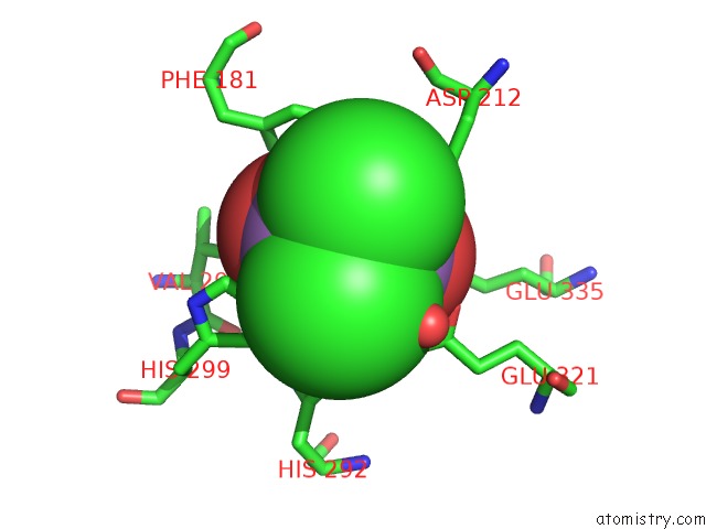
Arsenic binding site 1 out of 3 in 5cnx
Go back to
Arsenic binding site 1 out
of 3 in the Crystal Structure of Xaa-Pro Aminopeptidase From Escherichia Coli K12

Mono view

Stereo pair view
Mono view
Stereo pair view
A full contact list of Arsenic with other atoms in the As binding
site number 1 of Crystal Structure of Xaa-Pro Aminopeptidase From Escherichia Coli K12 within 5.0Å range:
|
Arsenic binding site 2 out of 3 in 5cnx
Go back to
Arsenic binding site 2 out
of 3 in the Crystal Structure of Xaa-Pro Aminopeptidase From Escherichia Coli K12
Mono view

Stereo pair view
Mono view
Stereo pair view
A full contact list of Arsenic with other atoms in the As binding
site number 2 of Crystal Structure of Xaa-Pro Aminopeptidase From Escherichia Coli K12 within 5.0Å range:
|
Arsenic binding site 3 out of 3 in 5cnx
Go back to
Arsenic binding site 3 out
of 3 in the Crystal Structure of Xaa-Pro Aminopeptidase From Escherichia Coli K12

Mono view

Stereo pair view
Mono view
Stereo pair view
A full contact list of Arsenic with other atoms in the As binding
site number 3 of Crystal Structure of Xaa-Pro Aminopeptidase From Escherichia Coli K12 within 5.0Å range:
|
Reference:
V.N.Are,
A.Kumar,
V.D.Goyal,
S.S.Gotad,
B.Ghosh,
R.Gadre,
S.N.Jamdar,
R.D.Makde.
Structures and Activities of Widely Conserved Small Prokaryotic Aminopeptidases-P Clarify Classification of M24B Peptidases Proteins 2018.
ISSN: ESSN 1097-0134
PubMed: 30536999
DOI: 10.1002/PROT.25641
Page generated: Mon Jul 7 00:13:06 2025
ISSN: ESSN 1097-0134
PubMed: 30536999
DOI: 10.1002/PROT.25641
Last articles
I in 3S2OI in 3RVI
I in 3S2H
I in 3RU1
I in 3S18
I in 3RX6
I in 3RSI
I in 3RG2
I in 3RTN
I in 3RTM