Arsenic »
PDB 5oc4-5yva »
5oc4 »
Arsenic in PDB 5oc4: Crystal Structure of Human Trna-Dihydrouridine(20) Synthase Dsrbd R361A-R362A Mutant
Protein crystallography data
The structure of Crystal Structure of Human Trna-Dihydrouridine(20) Synthase Dsrbd R361A-R362A Mutant, PDB code: 5oc4
was solved by
C.Bou-Nader,
L.Pecqueur,
D.Hamdane,
with X-Ray Crystallography technique. A brief refinement statistics is given in the table below:
| Resolution Low / High (Å) | 41.97 / 1.71 |
| Space group | H 3 |
| Cell size a, b, c (Å), α, β, γ (°) | 83.940, 83.940, 56.500, 90.00, 90.00, 120.00 |
| R / Rfree (%) | 19.4 / 21.5 |
Other elements in 5oc4:
The structure of Crystal Structure of Human Trna-Dihydrouridine(20) Synthase Dsrbd R361A-R362A Mutant also contains other interesting chemical elements:
| Chlorine | (Cl) | 1 atom |
Arsenic Binding Sites:
The binding sites of Arsenic atom in the Crystal Structure of Human Trna-Dihydrouridine(20) Synthase Dsrbd R361A-R362A Mutant
(pdb code 5oc4). This binding sites where shown within
5.0 Angstroms radius around Arsenic atom.
In total only one binding site of Arsenic was determined in the Crystal Structure of Human Trna-Dihydrouridine(20) Synthase Dsrbd R361A-R362A Mutant, PDB code: 5oc4:
In total only one binding site of Arsenic was determined in the Crystal Structure of Human Trna-Dihydrouridine(20) Synthase Dsrbd R361A-R362A Mutant, PDB code: 5oc4:
Arsenic binding site 1 out of 1 in 5oc4
Go back to
Arsenic binding site 1 out
of 1 in the Crystal Structure of Human Trna-Dihydrouridine(20) Synthase Dsrbd R361A-R362A Mutant
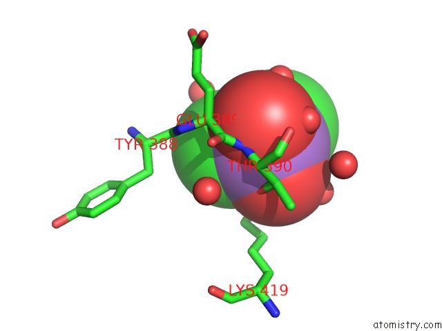
Mono view
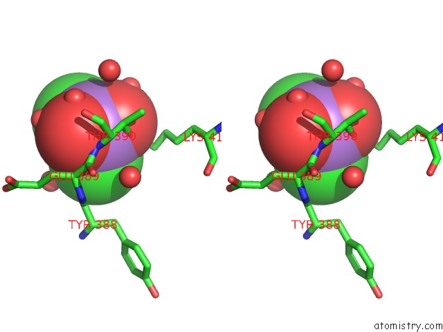
Stereo pair view
Mono view
Stereo pair view
A full contact list of Arsenic with other atoms in the As binding
site number 1 of Crystal Structure of Human Trna-Dihydrouridine(20) Synthase Dsrbd R361A-R362A Mutant within 5.0Å range:
|
Reference:
C.Bou-Nader,
P.Barraud,
L.Pecqueur,
J.Perez,
C.Velours,
W.Shepard,
M.Fontecave,
C.Tisne,
D.Hamdane.
Molecular Basis For Transfer Rna Recognition By the Double-Stranded Rna-Binding Domain of Human Dihydrouridine Synthase 2. Nucleic Acids Res. V. 47 3117 2019.
ISSN: ESSN 1362-4962
PubMed: 30605527
DOI: 10.1093/NAR/GKY1302
Page generated: Mon Jul 7 00:28:15 2025
ISSN: ESSN 1362-4962
PubMed: 30605527
DOI: 10.1093/NAR/GKY1302
Last articles
Mg in 3T2CMg in 3T2B
Mg in 3T1R
Mg in 3T1O
Mg in 3T0D
Mg in 3T1Q
Mg in 3T12
Mg in 3T1K
Mg in 3T10
Mg in 3T0Z